
- Home page
- buying guide
- mammography unit
mammography unit
mammography unit: A technical guide
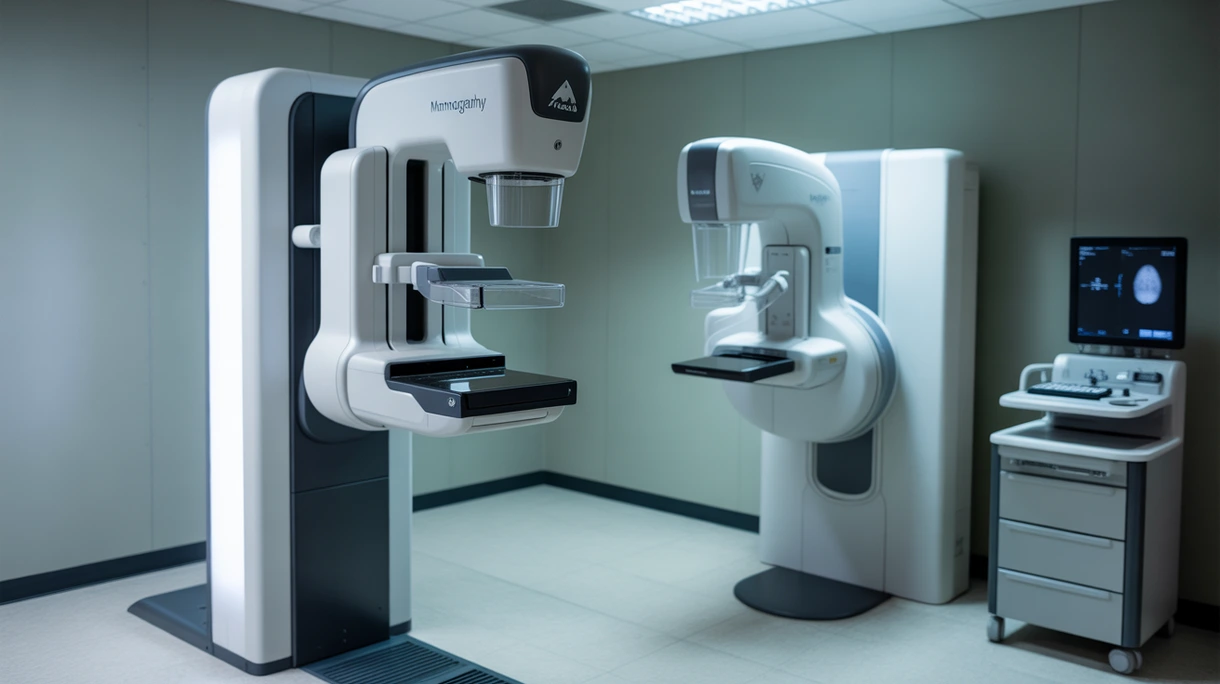
4 Criteria to choose your mammography unit
Image Spatial Resolution
High pixel pitch ensures detection of microcalcifications and subtle architectural distortions vital for early stage breast cancer diagnosis accuracy.
Average Glandular Dose
Automated exposure control systems optimize radiation levels to maintain high signal-to-noise ratios while strictly adhering to safety protocols.
Dynamic Compression Control
Intelligent paddles provide consistent pressure across varied tissue densities to minimize patient discomfort and prevent motion artifacts during acquisition.
DICOM Connectivity Compliance
Seamless integration with existing archive and information architectures ensures efficient diagnostic reporting and facilitates secure longitudinal patient data management.
Available technologies and advantages
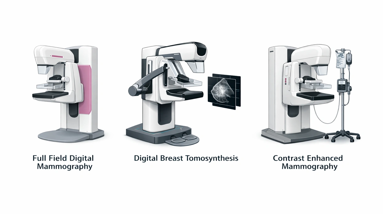
Full Field Digital Mammography
FFDM utilizes solid-state detectors to convert X-rays into electrical signals providing superior contrast compared to traditional screen-film systems while optimizing diagnostic workflow. These units offer rapid throughput and reliable imaging for screening programs but lack the three-dimensional depth needed for dense tissue.
Digital Breast Tomosynthesis
DBT captures multiple low-dose projections from different angles to reconstruct a three-dimensional volume for comprehensive tissue layer analysis and accurate diagnostic visualization. This technology significantly reduces false positives and improves cancer detection rates although it requires higher storage capacity and reading time.
Contrast Enhanced Mammography
Dual-energy imaging combined with intravenous iodine contrast agents highlights areas of neovascularization to differentiate between benign and malignant lesions for surgical planning. It serves as a cost-effective alternative to MRI for surgical planning despite the added risk of patient contrast reactions.
6 expert tips
Prioritize systems featuring wide-angle tomosynthesis to maximize the detection of invasive cancers in patients with dense breast tissue.
Invest in platforms offering synthetic two-dimensional imaging to eliminate the need for additional radiation during three-dimensional tomosynthesis examinations.
Select units equipped with ergonomic paddle designs to increase patient comfort and improve screening compliance across diverse clinical populations.
Evaluate the long-term total cost of ownership including detector replacement frequency and annual software maintenance for budget sustainability.
Ensure the imaging software includes advanced detection tools to assist radiologists in identifying suspicious lesions with high diagnostic sensitivity.
Confirm the presence of high-resolution monitors to ensure that the primary diagnostic workstation meets rigorous medical display standards.
Always verify the technical certification to ensure compliance with local building safety standards and long-term durability.
Innovations and Trends
AI-driven lesion prioritization reduces radiologist workload while improving diagnostic accuracy and consistency across complex screening populations and varied protocols.
This emerging technology measures individual X-ray photons to provide unprecedented spatial resolution and energy-weighted images at lower doses.
Combined modality platforms offer simultaneous anatomical and morphological assessment to enhance diagnostic confidence for patients with extremely dense breasts.
Cloud-based quality control systems enable real-time monitoring of detector performance to ensure continuous compliance with strict regulatory imaging standards.
*Prices are pre-tax. They exclude delivery charges and customs duties and do not include additional charges for installation or activation options. Prices are indicative only and may vary by country, with changes to the cost of raw materials and exchange rates.
About MedicalExpo
MedicalExpo is a marketplace that offers B2B players a complete experience, from product sourcing to purchase. MedicalExpo is an entity of VirtualExpo Group.
Are you a buyer?
Are you a supplier?