- Primary care
- General practice
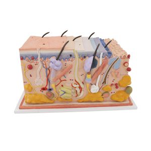
- Skin anatomical model
- 3B Scientific
Skin anatomical model J15for teachingfor cancers
Add to favorites
Compare this product
Characteristics
- Area of the body
- skin
- Procedure
- for teaching
- Simulated pathology / condition
- for cancers
Description
This 3B Scientific® Skin Pathology model shows healthy skin and 5 different stages of malignant melanoma on the front and back, enlarged 8 times:
healthy
malignant cells are found at the surface, within the epidermis
malignant cells fill the epidermis, a few invade the papillary layer
malignant cells fill the papillary layer
malignant cells invade the reticular layer
malignant cells have reached the subcutaneous fatty tissue, satellite cells approach a vein
Catalogs
Exhibitions
Meet this supplier at the following exhibition(s):

Related Searches
- 3B Scientific anatomical model
- 3B Scientific training anatomical model
- 3B Scientific teaching anatomical model
- Stethoscope
- 3B Scientific bone model
- 3B Scientific flexible anatomical model
- 3B Scientific skull model
- 3B Scientific tooth model
- 3B Scientific plastic anatomical model
- Dental anatomical model
- 3B Scientific mouth anatomical model
- Vascular model
- 3B Scientific body anatomical model
- Training vascular model
- White anatomical model
- Leg anatomy model
- 3B Scientific spine anatomical model
- Digestive system model
- 3B Scientific pelvis model
- Circulatory system vascular model
*Prices are pre-tax. They exclude delivery charges and customs duties and do not include additional charges for installation or activation options. Prices are indicative only and may vary by country, with changes to the cost of raw materials and exchange rates.


