

- Aesthetic medicine, Wellness
- Aesthetic medicine
- Skin melanin analysis system
- Beijing Sanhe Beauty


- Company
- Products
- Catalogs
- News & Trends
- Exhibitions
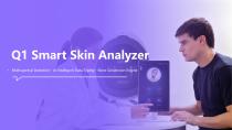
Skin imaging system Q1 seriesskin conditionskin vascularizationskin pigmentation


Add to favorites
Compare this product
Characteristics
- Type of diagnosis
- skin imaging, skin condition, skin vascularization, skin pigmentation, cutaneous sebum, skin colorimetry, UV skin imaging, skin topography, melanin level
Description
Overview
Skin analysis device providing multi-spectral imaging and AI-assisted detection for a comprehensive assessment of facial skin condition in clinical dermatology, aesthetic and professional skincare settings.
Technology
Eight spectral imaging capturing eight distinct image sets; high-definition industrial camera; AI algorithms for feature extraction, enhancement and diagnostic map generation.
Indication
The system is intended to analyze skin color status, wrinkles, pores, pigmentation, acne, sub-epidermal uneven tones and microvascular structures, sebum secretion and deep acne, flora distribution, sensitive zones and other skin conditions by combining multiple imaging spectra with AI processing for objective clinical assessment.
Imaging modes and functions
Analysis & Output
AI-driven detection and dimensional parsing with generation of diagnostic maps (red map, thermal/heat map) to visualize sensitivity, inflammation and blood distribution; enhanced recognition of skin texture and clinical markers for documentation and treatment planning.
Imaging hardware
Integrated high-definition industrial camera and controlled illumination system for reproducible captures and reliable image quality.
Technical specifications
Skin analysis device providing multi-spectral imaging and AI-assisted detection for a comprehensive assessment of facial skin condition in clinical dermatology, aesthetic and professional skincare settings.
Technology
Eight spectral imaging capturing eight distinct image sets; high-definition industrial camera; AI algorithms for feature extraction, enhancement and diagnostic map generation.
Indication
The system is intended to analyze skin color status, wrinkles, pores, pigmentation, acne, sub-epidermal uneven tones and microvascular structures, sebum secretion and deep acne, flora distribution, sensitive zones and other skin conditions by combining multiple imaging spectra with AI processing for objective clinical assessment.
Imaging modes and functions
- RGB white light: natural, soft and uniform illumination with multi-directional diffused light to document skin color, wrinkles, pores, pigmentation and acne; serves as a reference for clinical assessment.
- Parallel polarized light: parallel polarized lens group to enhance surface reflection, improving observation of skin texture, fine lines and wrinkles; supports AI wrinkle-counting algorithms.
- Cross polarized light: cross polarization to attenuate surface reflection and reveal sub-epidermal uneven tone and hidden microvascular structures.
- UV light: 365 nm wavelength with filter to reveal epidermal pigmentation, pigment residues of acne scars and deep pigmentation from UV exposure.
- Blue light: 405 nm wavelength with filter; uses skin fluorescence to visualize sebum secretion, deep acne and microbial flora distribution.
- Wood's (Wu's) light: WOOD'S mode to observe subcutaneous vascular and pigmented lesions, brown spots, erythema and to detect fluorescence from topical agents or cosmetics.
- Composite / Erythromin light: combined spectrum with AI to produce a red map highlighting blood distribution and deep inflammation for sensitivity and redness assessment.
- Composite / Thermal light: combined spectrum plus AI to generate a heat/thermal map indicating reactive or inflamed areas with elevated hemoglobin signal.
Analysis & Output
AI-driven detection and dimensional parsing with generation of diagnostic maps (red map, thermal/heat map) to visualize sensitivity, inflammation and blood distribution; enhanced recognition of skin texture and clinical markers for documentation and treatment planning.
Imaging hardware
Integrated high-definition industrial camera and controlled illumination system for reproducible captures and reliable image quality.
Technical specifications
- Eight spectral imaging: captures eight distinct spectral image sets.
- UV imaging wavelength: 365 nm (with filter) for epidermal pigmentation analysis.
- Blue light wavelength: 405 nm (with filter) for fluorescence-based sebum and flora visualization.
- Polarized imaging modes: parallel and cross-polarized lens groups for surface and subsurface feature enhancement.
- AI-assisted analysis: wrinkle counting, red/thermal mapping, sensitivity and inflammation detection.
- High-definition industrial camera for image acquisition and clinical documentation.
- Available visualizations: RGB white light, polarized lights, UV, blue, Wood's mode, composite/erythromin and composite/thermal outputs.
- Outputs: multi-spectral image sets and AI-generated diagnostic maps (red map, heat/thermal map).
VIDEO
Catalogs
Skin imaging system Q1 Series
29 Pages
Other Beijing Sanhe Beauty products
Skin Analysis


Related Searches
- SANHE aesthetic medicine laser
- SANHE skin and body treatment unit
- SANHE trolley-mounted laser
- SANHE diode laser
- Tabletop laser
- SANHE dermatology laser
- SANHE hair removal laser
- SANHE Nd:YAG laser
- SANHE skin rejuvenation laser
- SANHE millisecond laser
- Surgical laser
- SANHE tattoo removal laser
- CO2 laser
- SANHE pigmented lesion treatment laser
- Acne treatment laser
- Microsecond laser
- SANHE picosecond laser
- Scar removal laser
- Vascular lesion treatment laser
- Nanosecond laser
*Prices are pre-tax. They exclude delivery charges and customs duties and do not include additional charges for installation or activation options. Prices are indicative only and may vary by country, with changes to the cost of raw materials and exchange rates.
About MedicalExpo
MedicalExpo is a marketplace that offers B2B players a complete experience, from product sourcing to purchase. MedicalExpo is an entity of VirtualExpo Group.
Are you a buyer?
Are you a supplier?