- Primary care
- General practice
- Nerve model
- Denoyer-Geppert

- Products
- Catalogs
- News & Trends
- Exhibitions
Body anatomical model 0167-00nerveneuronfor teaching
Add to favorites
Compare this product
fo_shop_gate_exact_title
Characteristics
- Area of the body
- body, nerve, neuron
- Procedure
- for teaching
Description
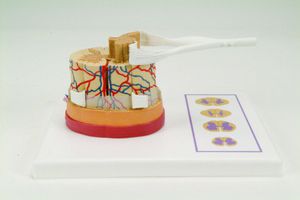
Magnified more than 2500 times and fully three-dimensional, this neuron model is depicted in its natural setting. With the membranous envelope cut away, the cytological ultrastructure, organelles and inclusions within the cell body are depicted in contrasting colors. A section of the axon lifts off to expose the enveloping myelin sheath and neuro-lemma, as well as the Schwann cell that formed them. Dendrites of the neuron extend into the background, and synaptic vesicles carrying neurotransmitters can be seen via a cutaway view. 54 features are identified in an illustrated key.
Nucleus
Nucleolus
Cytoplasm
Lysosome
Vesicles (emerge from Golgi & ER)
Golgi apparatus of Golgi complex
Mitochondria
Endoplasmic reticulum (ER) Nissl body
(rough ER has polyribosomes on it)
Nerve bers
Axon hillock (cone-shaped base of axon)
Axon
Dendrite
Schwann cell cytoplasm
Node of Ranvier
Cytoplasm of axon (axoplasm) includes:
neuro laments, microtubules, vesicles and
mitochondria
Axolemma
Schwann cell nucleus
Schmidt-Lanterman cleft
Myelin sheath (wrapping portion with no
cytoplasm)
Related Searches
- Anatomy model
- Demonstration anatomical model
- Teaching anatomy model
- Bone anatomical model
- Flexible anatomical model
- Intracranial anatomical model
- Denture model
- Plastic anatomy model
- Transparent anatomical model
- Oral anatomical model
- Whole body anatomical model
- Leg anatomy model
- Vertebral column model
- Digestive system model
- Pelvic anatomical model
- Cardiac anatomical model
- Nervous system model
- Facial model
- Articulated anatomical model
- Circulatory system model
*Prices are pre-tax. They exclude delivery charges and customs duties and do not include additional charges for installation or activation options. Prices are indicative only and may vary by country, with changes to the cost of raw materials and exchange rates.


