
- Products
- Catalogs
- News & Trends
- Exhibitions
Analysis software module MYRIAN® XP-BRAINmanagementvisualizationdiagnostic
Add to favorites
Compare this product
fo_shop_gate_exact_title
Characteristics
- Function
- analysis, management, visualization, diagnostic, evaluation, treatment, navigation
- Applications
- CT, neurological
- Area of the body
- head
Description
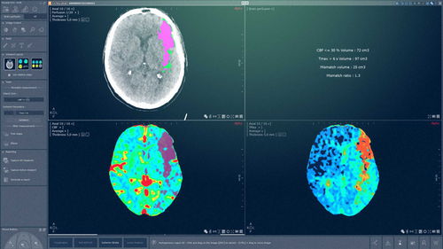
This XP-Brain Perfusion CT module aims to create, visualize, and analyze parametric maps allowing the study of the hemodynamic parameters of the cerebral parenchyma based on specifically acquired series, so-called cerebral perfusion series.
The analysis of these parametric maps is carried out in the following clinical context: provide information that can be used to help to take decisions with therapeutic purpose in case of a diagnosed cerebral ischemia linked to a deficit in vascularization.
Application
Dedicated layout and workflow for ischemic lesion
Automatic motion correction
Automatic detection of cerebral scythe
Automatic segmentation of brain parenchyma and ventricles
Automatic calculation of local AIF and VOF
Automatic computation of perfusion results (CBV, CBF, rCBF, Tmax, MTT, TTP)
Automatic computation of quantified and color-coded CT Perfusion maps
Dedicated correction tools for each automatic process
Automatic computation of mismatch volume and ratio
Correction tools of mismatch analysis (modification rCBF and Tmax parameters)
Symmetric ROI measurements
Dedicated report
Catalogs
No catalogs are available for this product.
See all of Intrasense‘s catalogsExhibitions
Meet this supplier at the following exhibition(s):

Related Searches
- Software module
- Analysis software module
- Clinical software module
- Radiology software module
- Viewer software module
- Management software module
- Measurement software module
- Scheduling software module
- Evaluation software module
- Diagnostic software module
- Surgical software module
- Automated software module
- Reporting software module
- Simulation software module
- Treatment software module
- Acquisition software module
- 3D simulation software module
- CT software module
- Radiography software module
- Tracking software module
*Prices are pre-tax. They exclude delivery charges and customs duties and do not include additional charges for installation or activation options. Prices are indicative only and may vary by country, with changes to the cost of raw materials and exchange rates.
