
- Company
- Products
- Catalogs
- News & Trends
- Exhibitions
Angiography software module VASCAN densitydata analysisvisualization

Add to favorites
Compare this product
Characteristics
- Applications
- angiography, density
- Function
- data analysis, visualization, scan, eye tracking, tracking
- Area of the body
- retina
- Type
- automated, AI-assisted
- Deployment mode
- for tablet PC
Description
Powered by Complex Optical Micro Angiography (COMAG) algorithm that uses phase and amplitude signal data to generate find quality OCTA images
AI-powered high-resolution OCTA imaging, offering superior clarity
Based on the powerful platform Mocean 4000 with reliable retinal tracking system, motion artitifacts are effectively minimized
Up to 12x8 mm widefield OCTA scan
Comprehensive data analysis including density, FAZ, imperfusion and flow analysis.
Up to 12x8mm widefield OCTA scan
VASCAN provides up to 12 x 8 mm extra wide angiography scan in a single shot, which gives you a overview of the the retinal vasculature within just a few seconds.
The AI-based algorithm enhances image clarity by eliminating projection artifacts, which results in improved visualization of the vessels.
Accurate Eye Tracking with SLO Imaging
SLO imaging serves as a dependable reference for an accurate eye tracker, effectively reducing artifacts, particularly in cases involving less cooperative patients.
Comprehensive OCTA analysis
The VASCAN OCTA module delivers high-resolution images featuring seven-layer segmentation, including VRI, superficial, intermediate, deep, outer retina, choriocapillaris, and choroid. It also offers robust data analysis capabilities, such as density analysis, FAZ analysis, impairment analysis, and flow area analysis, ensuring comprehensive insights.
Enhanced vessel clarity in choroidal OCTA
Thanks to the enhanced algorithm, choroidal OCT angiography imaging now provides a clearer view of the vessels.
Catalogs
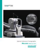
Mocean™ 4000
8 Pages
Other Moptim products
Diagnostic


*Prices are pre-tax. They exclude delivery charges and customs duties and do not include additional charges for installation or activation options. Prices are indicative only and may vary by country, with changes to the cost of raw materials and exchange rates.