
Cloud-Based Medical Image Management Image Viewing and Archiving eNcounterCloud® is a secure, cloud-based, web application for medical image archiving ...
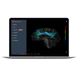
Review all automatically computed DSC maps. Compare findings through AI-powered ROI statistics. Export maps and core findings to your PACS instantly. DTI Single-click Fiber Tracking. Accelerate your DTI post-processing workflow ...

... module - enables creating panoramic image of several constituent images of eye fundus. - Optic nerve head module - serves to calculate proportions between the area of optic disc cup and its total area. - ...
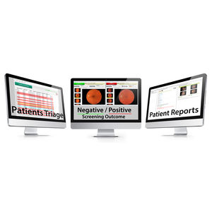
... EyeArt™ is a fully automated, CE marked DR screening software. EyeArt™ is an Artificial Intelligence software platform featuring cutting–edge image analysis and classification ...
Eyenuk, Inc.
Simpleware 3D image processing and modeling software is your high-end solution for working with medical image data. Intuitive tools and filters allow you to accurately ...
Synopsys Simpleware Product Group
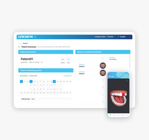
... information, treatment report Multi-color analysis Analyze MCA map of captured teeth Examination Reports Select and transmit only necessary information instantly View in 3D View 360-degree rotating ...
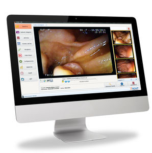
Designed to work with any endoscopy equipment, Zscan Software is the ideal tool for image managing, reporting and results delivery. Our software is present in every step of your medical ...
Zscan software

... edit and apply effects to multiple photos simultaneously on a single screen. Its intuitive assistive features make aligning images both simple and efficient. Align everything & Make your own standard On a single ...
PhotoAlign

... visible in the image Segmentation of the wound area after defining a starting point Calculation of the area and extent of the wound taking into account the geometric distortion of the ruler Benefits: Expertise ...
softgate gmbh

... to process ultrasound images for sonographic characteristics, helping them to make more confident diagnostic decisions. Our platform uses statistical pattern recognition and quantification methods ...
AmCad BioMed

... Florida Probe software is the core of both our Florida Probe and VoiceWorks Systems. Yes, it is periodontal charting software, but also so much more! What Does It Do? Sounds and Images ...

... earlier precision on the frontlines of CNS disease. QyScore® is a groundbreaking neuroimaging analysis platform for both clinical routine and clinical trial settings. Our technology is ...

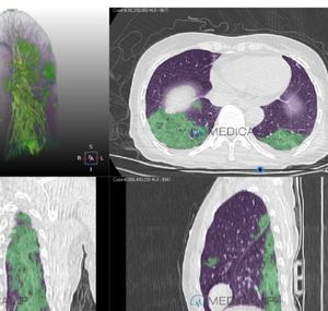
... Suitable for the screening, diagnose and analyze of adult chest CT images. Provides identification and alert for chest fracture. Supports MPR, bone VR, rib analysis, deep learning image ...
... position • Various image export tools(CD, DVD, Print) • DICOM compatible Advanced Image Processing technology and Display Tools • Improved diagnostic accuracy through image ...
Through deep learning technology, a CT image of the patient’s pneumonia lesions caused by COVID-19 can be automatically detected, segmented and quantified in about 1 minute. The proportion of the lesion area within the ...

DentiqRecon features special reconstruction modes that enhance the image quality while reducing a variety of CT artifacts caused by beam-hardening and scattering effects.

BU-CADTM is a software application indicated to assist trained interpreting physicians in analyzing the breast ultrasound images of patients with soft tissue breast lesions suspicious for breast cancer ...
... the soft ware for Digiray FireCR Dental PSP Readers. Experience the high diagnostic value of your FireCR Dental PSP Reader. Aft er just a few seconds of scanning, the image appears. The chronological ...
Digiray

... Exudates, Soft Exudates, Hemorrhages, Microaneurysms) Resolution Requirement 800*800 Fundus Camera Works with MiiS AI-DR Software Brand CenterVue Optovus Canon Topcon ZEISS Nidek Miis

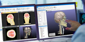
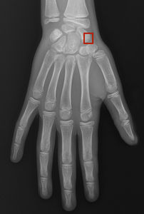
... Axis Deviation ● Femur length ● Tibia length ● Full leg length ● Length difference between legs (for bilateral images in the absence of THA) Benefits Automated Quicker pre-selection by instantly ...
Imagebiopsy Lab
Your Artificial Intelligence Partner on X-rays 27% time saved 67% False negatives avoided 99% Sensitivity 89% Specificity Exams sorted by priority order Rayvolve detects and automatically ranks X-Rays with abnormalities, so ...
AZmed

... Why Duali-Q? AI automation Our AI automatically analyzes radiology images to detect commonly-overlooked findings, such as pulmonary nodules and liver lesions, comparing image data with report text, ...

Medmont Studio is our proprietary software integrating the medmont meridia Advanced Topographer, E300 Corneal Topographer, Medmont M700 Automated Perimeter, and DV2000 Imaging Software together in a unified ...
Medmont

The VF Sync software has been developed to connect multiple ProEX Telehealth Hub units over a local or wide area network and to facilitate the integration with the customers electronic health record software. ...
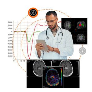
Our breakthrough neuroimaging tools seamlessly integrate into your daily clinical CT & MRI workflows to assist you in every step of your decision-making in pre- and post-treatment environments. Seamless neuroimaging ...
Olea Medical
Please specify:
Help us improve:
remaining