


{{product.productLabel}} {{product.model}}
{{#if product.featureValues}}{{product.productPrice.formattedPrice}} {{#if product.productPrice.priceType === "PRICE_RANGE" }} - {{product.productPrice.formattedPriceMax}} {{/if}}
{{#each product.specData:i}}
{{name}}: {{value}}
{{#i!=(product.specData.length-1)}}
{{/end}}
{{/each}}
{{{product.idpText}}}
{{product.productLabel}} {{product.model}}
{{#if product.featureValues}}{{product.productPrice.formattedPrice}} {{#if product.productPrice.priceType === "PRICE_RANGE" }} - {{product.productPrice.formattedPriceMax}} {{/if}}
{{#each product.specData:i}}
{{name}}: {{value}}
{{#i!=(product.specData.length-1)}}
{{/end}}
{{/each}}
{{{product.idpText}}}
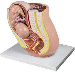

... This life size model depicts the female human pelvis in median section with a fetus in the 32nd week of pregnancy. The fetus is in the normal presentation and position. The model depicts graphically the ...
Erler-Zimmer

Weight: 0.9 kg
... Natural casting of an adult female pelvis. The sacrum is removable and the movements in the iliosacral joint can be demonstrated. ...
Erler-Zimmer
Length: 22 cm
Width: 20 cm
Height: 25 cm
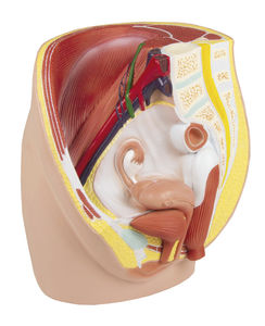
... This life size model is median sagittal sectioned and shows the female pelvis in 3 parts. Additional to muscles and vessels of the abdomen it depicts particularly the urogenital organs. Removable are one half of the uterus, ...
Erler-Zimmer
Length: 21 cm
Width: 19 cm
Height: 25 cm
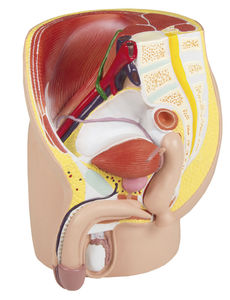
... This life size model is median sagittal sectioned and shows the male pelvis in 4 parts. Additional to muscles and vessels of the abdomen it depicts particularly the urogenital organs. Removable are one half of the cavernous ...
Erler-Zimmer

... This 3D print displays a male left pelvis and proximal thigh, sectioned midsagittally and transversely through L3/4. It highlights superficial and deep structures of the pelvis, inguinal region, and proximal thigh. Muscles ...
Erler-Zimmer

... printed specimen displays the lower posterior abdominal wall, pelvic cavity, and proximal thigh, including the gluteal regions and femoral triangles. Lower Posterior Abdominal Wall & False Pelvis Transected at L2/L3, ...
Erler-Zimmer

... This 3D printed specimen displays superficial and deep structures of the pelvis, inguinal region, femoral triangle, and gluteal region, offering a detailed view of female pelvic anatomy. Posterior Abdominal Wall & ...
Erler-Zimmer
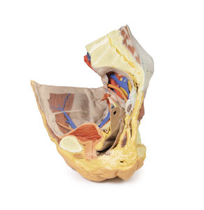
... uterus, and lateral pelvic wall, while the obturator nerve, artery, and vein pass through the obturator canal. Sacral nerves (S1–S3) form the sciatic nerve, which exits via the greater sciatic foramen alongside gluteal vessels. Pelvic ...
Erler-Zimmer
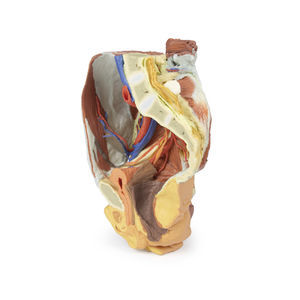
... This 3D printed female right pelvis is sectioned midsagittally and transversely at L4 and the proximal thigh, revealing the true and false pelvis, inguinal region, femoral triangle, and gluteal region. Muscles & Nerves Key ...
Erler-Zimmer
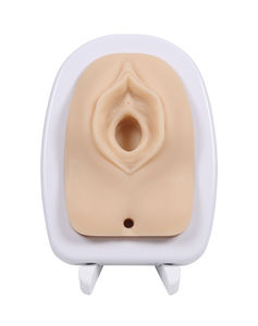
Weight: 0.7 kg
This representation of a female vulva with clitoris, labia, urethral opening, vaginal opening, anus, perineum and muscles is made of reinforced material and allows you to practise suturing birth injuries realistically over and over again. The muscles ...
Erler-Zimmer