


{{product.productLabel}} {{product.model}}
{{#if product.featureValues}}{{product.productPrice.formattedPrice}} {{#if product.productPrice.priceType === "PRICE_RANGE" }} - {{product.productPrice.formattedPriceMax}} {{/if}}
{{#each product.specData:i}}
{{name}}: {{value}}
{{#i!=(product.specData.length-1)}}
{{/end}}
{{/each}}
{{{product.idpText}}}
{{product.productLabel}} {{product.model}}
{{#if product.featureValues}}{{product.productPrice.formattedPrice}} {{#if product.productPrice.priceType === "PRICE_RANGE" }} - {{product.productPrice.formattedPriceMax}} {{/if}}
{{#each product.specData:i}}
{{name}}: {{value}}
{{#i!=(product.specData.length-1)}}
{{/end}}
{{/each}}
{{{product.idpText}}}


... new Augmented Anatomy App in combination with this high quality anatomical model makes learning now even easier and more efficient!This app automatically recognises our anatomical models ...
Erler-Zimmer

Weight: 0.9 kg
Natural casting of an adult female pelvis. The sacrum is removable and the movements in the iliosacral joint can be demonstrated.
Erler-Zimmer

... this very successful model can be offered at a very reasonable price, so everyone can afford this extraordinary model. Augmented- Anatomy- App The new Augmented Anatomy App in combination with this high quality ...
Erler-Zimmer

Length: 23 cm
Width: 16.5 cm
Height: 17 cm
... muscle, temporal muscle, pterygoideus medialis and lateralis muscles) are represented in the form of elastic bands. Using this model, it is possible to demonstrate the function of the masticatory muscles with closure of the mandible, ...
Erler-Zimmer

Length: 19 cm
Width: 14 cm
Height: 43 cm
... Approximately half-size representation of the human torso. Ideal for use as a desk model or for studying basic anatomy. The model consists of the following parts: - Base body, two halves of the head - One with ...
Erler-Zimmer

... This 3D print displays a male left pelvis and proximal thigh, sectioned midsagittally and transversely through L3/4. It highlights superficial and deep structures of the pelvis, inguinal region, and proximal thigh. Muscles and Fascia The transverse ...
Erler-Zimmer

... This multipart 3D printed specimen displays the lower posterior abdominal wall, pelvic cavity, and proximal thigh, including the gluteal regions and femoral triangles. Lower Posterior Abdominal Wall & False Pelvis Transected at L2/L3, it shows the ...
Erler-Zimmer

... This 3D printed specimen displays superficial and deep structures of the pelvis, inguinal region, femoral triangle, and gluteal region, offering a detailed view of female pelvic anatomy. Posterior Abdominal Wall & Spine Sectioned at L4, it shows ...
Erler-Zimmer
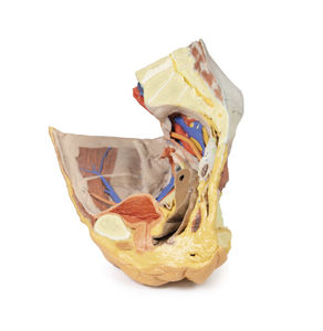
... This 3D printed specimen preserves superficial and deep structures of the true and false pelvis, the inguinal ligament, obturator canal, and sciatic foramina, with ‘windows’ exposing extraperitoneal structures. Posterior Abdominal Wall & Major Vessels Sectioned ...
Erler-Zimmer
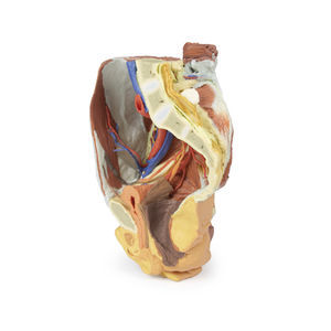
... This 3D printed female right pelvis is sectioned midsagittally and transversely at L4 and the proximal thigh, revealing the true and false pelvis, inguinal region, femoral triangle, and gluteal region. Muscles & Nerves Key muscles include psoas, ...
Erler-Zimmer