
{{product.productLabel}} {{product.model}}
{{#if product.featureValues}}{{product.productPrice.formattedPrice}} {{#if product.productPrice.priceType === "PRICE_RANGE" }} - {{product.productPrice.formattedPriceMax}} {{/if}}
{{#each product.specData:i}}
{{name}}: {{value}}
{{#i!=(product.specData.length-1)}}
{{/end}}
{{/each}}
{{{product.idpText}}}

Length: 22 cm
Width: 18 cm
Height: 46 cm
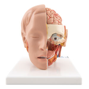
This high quality model represents the outer, superficial and the internal (median section) structures of head and neck. The half head with musculature is delivered on removable stand ...
3B Scientific

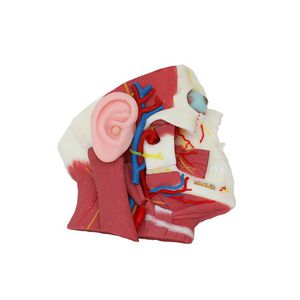
The left half of this life-size model in midsagittal section shows the muscles, with nerves, vessels and bony structures and contains a removable brain half. The head is mounted on a detachable neck part ...
3B Scientific
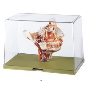
Our most detailed head model! This life-size 6-part head is mounted on a base and features a removable 4-part brain half with arteries. The eyeball with optic nerve is also removable ...
3B Scientific
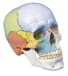
Skull model like ref.no. 4500, additionally with didactical painting of individual bones on one side of the skull. The bones are numbered referring to the included nomenclature. Size: 18 x 19 x 12 cm, weight: 0.7 kg
Erler-Zimmer

This skull model is an actual cast of a real human specimen and shows all anatomical structures in highest detail. It is made for students in anatomy, medicine, surgery, otolaryngology, ophthalmology and dentistry. The ...
Erler-Zimmer

Length: 15 cm
Width: 15 cm
Height: 30 cm
This life size head model is dissected along the sagittal plane into 2 halves. Details of the oronasal cavity and larynx as well as musculature of the pharynx are exceptionally well represented. Mounted ...
Erler-Zimmer
Deep structure of inferior parotid gland soft silicone anatomy model Material: Environmental protection soft silicone rubber, environmental protection paint; The silicone rubber and its auxiliary chemical products ...
MeiWo Science

Posterior pharyngeal view and coronal section of face soft silicone anatomy model Material: Environmental protection soft silicone rubber and paint; The silicone rubber and its auxiliary chemical products ...
MeiWo Science

Soft Silicone Nasopharyngeal and Trachea Anatomy Model 1. Material: Environmental protection food grade soft silicone 2. Size: Natural 3. Parts: 2 parts 4. Teaching contents: The model mainly ...
MeiWo Science
Complete child dental manikin consisting of rod with joint, face mask that allows the use of water, which comes out of the manikin through a special drain in the back, skull cap, metal articulator with block in the maximum ...

Complete dental manikin consisting of rod with joint, face mask that allows the use of water, which comes out of the manikin through a special drain in the back, skull cap, metal articulator with block in the maximum ...

Neuro Vascular System Ⅸ Model mainly includes the vascular, aortic arch, femoral artery and intracranial, each part can be replaced with the corresponding lesion shape. It can be used to simulate femoral artery and radial ...
Trando 3D Medical Technology

Length: 650 mm
Width: 280 mm
... Cava Heart Model I Model: XX001J Material:Silicone Custom service:providing free design service&technical solutions Payment:T/T Lead time:7-15 days Port:Ningbo,China Shipping Methods:FedEx, DHL, ...
Trando 3D Medical Technology

ERCP Simulator I Model is 3D printed based on real CT data. It includes head, esophagus, stomach, duodenum and bile duct. Duodenum, bile duct and gallbladder are replacable. Three types of duodenal ...
Trando 3D Medical Technology
Length: 9 in
Width: 4 in
Height: 3 in
... Features: Textured surface with pancreatic notch, head, body and uncinate process. Typical Uses: These organs are used in the SynDaver Synthetic Human product line. They are also incorporated into complex model ...

This is the most complete solution to take your bucomaxillo facial surgery training to a new level. Buy this exclusive package now and have the best products on the market for your hands-on. 1X MANNEQUIN CLASS II ...

... are situated on a stand. Exhibited externally and internally on the left side are the arteries (colored red) supplying the head and neck, while on the right, the 12 cranial nerves (colored yellow) and the distribution ...
Denoyer-Geppert
Width: 32 cm
Height: 21 cm
Weight: 1.1 kg
Natural size, in SOMSO-Plast®. Showing the muscles, nerves and vessels, in particular trigeminal nerve and facial nerve. The tongue is removable. Separates into 2 parts. On a stand with green base, under a removable transparent ...
Coburger Lehrmittelanstalt
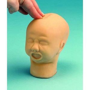
A robust life size foetal head designed for practising palpation of the skull through the skin to identify sutures and fontanelles. Comprises an outer flexible skin with an inner hard bone-like structure. Individual bones ...
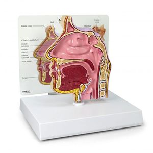
Full size cut-away normal model depicts a near median section through the nose and nasal passages. Details include nasal cavity, soft and hard palate, uvula, eustachian tube and pharyngeal tonsil. Reverse side shows ethmoid ...

Length: 30 cm
Width: 19 cm
Height: 42 cm
... transparent stickers with printed inscriptions can be removed from the model for learning purposes. This makes the model ideal for exam preparation. PRODUCT DETAILS Life size model Colour: ...

Length: 820 mm
Width: 440 mm
Height: 350 mm
... patient (head and torso). The head is made of a synthetic material that visually and palpationally imitates the skin. For realism of conducting practical exercises nose, eyes, ears and open mouth are ...
SATC solution

Basic yet comprehensive training model Soft type replaceable gingiva – 2 types to choose from (pink, pink for silicone impression taking) A wide variety of operative training is possible A wider facial ...

... male and female genitalia. The female organs include a fetus in the womb. Dissected into 23 parts: torso, female breast plate, head, eyeball, brain, vertebra spinal nerves, lung (2 parts), heart (2 parts), liver, kidney, ...
Xincheng Scientific Industries Co., Ltd.
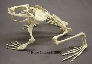
From West Africa’s cascading mountain streams, this is the largest frog in the world. The Goliath Frog, weighing over 7 pounds, with a body length of 12 1/2 inches (measuring over 3 feet from nose to toe) has a ten foot leap. This ...

Length: 33 cm
Width: 23 cm
Height: 5.5 cm
Life size representation of the superficial and the internal structure of the head and neck. This relief models shows all relevant structures of the human head in great detail.
Yuan Technology

This high quality classic skull is medically detailed in structure. It is the perfect tool for professional teaching and study of human anatomy. It can be disassembled into 3-part: Calvaria, Base of Skull, and Mandible. The calvaria is ...
Wellden International Inc.

The sagittal section of human head shows internal structures such as sinus and upper respiratory tract. The model is accompanied by a patient education card.

Face Mark with Drainage is a silicone face for the dental simulation head mold, which is easy to operate, feels real, and is soft and realistic. Description : Used in Dental Schools ...
Tangshan UMG Medical Instrument

EEG Teaching Head Model Position mark Plastic material
Please specify:
Help us improve:
remaining