
{{product.productLabel}} {{product.model}}
{{#if product.featureValues}}{{product.productPrice.formattedPrice}} {{#if product.productPrice.priceType === "PRICE_RANGE" }} - {{product.productPrice.formattedPriceMax}} {{/if}}
{{#each product.specData:i}}
{{name}}: {{value}}
{{#i!=(product.specData.length-1)}}
{{/end}}
{{/each}}
{{{product.idpText}}}
Length: 22 cm
Width: 10 cm
Height: 10 cm
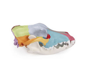
This life-size replica of a dog's skull shows the anatomical structures and is didactically painted so that the individual bones of the skull can be clearly seen and their location and relationship to one another can ...

Length: 18 cm
Width: 16 cm
Height: 22 cm
... optic nerve and blood vessels, the model eye is ideally suited for training and study as well as for patient information. The eye model is also ideal as a beautiful inventory in the veterinary ...
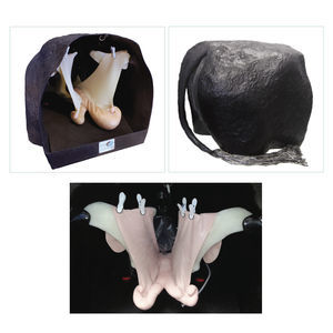
• Rear portion of our Hereford model constructed in epoxy/fiberglass • Constructed with pelvis, soft perineum palpation panel, inflatable vinyl rectum, and flexible tail. • Includes the Bovine Theriogenology Uterus ...
Veterinary Simulator Industries

... placentomes • Bovine Palpation Panel Assembly • Enables compatibility of our Bovine • Bovine Ovaries Set All models have representations of the cervix, broad ligament, and ovaries The Uterus Set includes ...
Veterinary Simulator Industries

Length: 51.2 in
Width: 13 in
Height: 24 in
Model to show anatomical skeleton of adult canine.

Length: 98.9 mm
... soft tissue as in 0906.1 Feline Hindlimb left with soft tissue Fracture - Intact model Size - medium Orientation - left Finishing - sand blasted Model Type - intact Structure - bone, ...
SYNBONE AG

Length: 325 mm
... patella, major ligaments and soft tissue Material - Solid foam Fracture - Intact model Size - medium Orientation - left Finishing - soft tissue with skin Model Type - intact Structure ...
SYNBONE AG
Please specify:
Help us improve:
remaining