
{{product.productLabel}} {{product.model}}
{{#if product.featureValues}}{{product.productPrice.formattedPrice}} {{#if product.productPrice.priceType === "PRICE_RANGE" }} - {{product.productPrice.formattedPriceMax}} {{/if}}
{{#each product.specData:i}}
{{name}}: {{value}}
{{#i!=(product.specData.length-1)}}
{{/end}}
{{/each}}
{{{product.idpText}}}

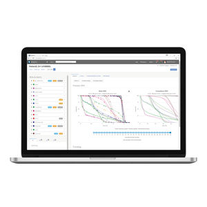
3mensio CT - Structural Heart & Endovascular Less invasive and more precise procedures with pre-op analysis Especially designed for and with cardiovascular specialists, 3mensio Structural Heart™ will let you plan aortic ...

... guided implant treatment is compatible with over 10,000 implants from more than 100 brands, as well as all DICOM compatible CT scanners and major optical and intraoral scanners. From dental scanning and planning, to ...
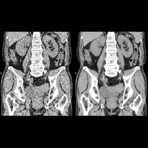
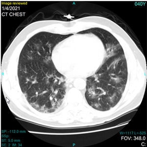
Sets a new direction in CT image quality with industry-leading low-contrast resolution and virtually noise*-free images. Innovations in hardware and the reconstruction algorithm result in a reconstruction speed – less ...
This function is for observing cross-sectional images of low-contrast regions, primarily tumor stains during procedures.

... best computer-aided software analysis tools Multi-disciplinary collaboration and online continued medical education frameworks We offer fully-fledged software applications* and OEM-facing complementary ...
VAREX Imaging

... efficient volume reconstruction is the hallmark of our 3D imaging software. Save time and avoid manual model conversion by simply loading your patient’s DICOM data from any cone beam CT machine. Invivo’s ...
Anatomage
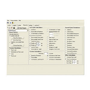
Collect data from a Brookfield CT3 Texture Analyzer and perform detailed data analysis including real time graphic plotting with TexturePro CT. Take control of your CT3 Texture Analyzer ...
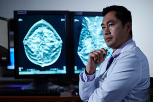
... faster procedure time, greater comfort, lower anxiety, and lower noise level.3 Expand Diagnostic Capabilities I-View CEM Software is a simple upgrade to any Selenia or 3Dimensions mammography system. Offering contrast ...
... risk. View dose distribution on planned as well as merged CT image sets. Compare daily and cumulative delivered dose to planned dose. Leverage Data with Automated Patient Dosimetry Software Monitor ...
Standard Imaging

The Planmed Verity® extremity CT scanner has an integrated touch screen with the Planmed Verity Manager software for operating the unit. Worklist management, imaging protocol optimization and image acquisition ...

... images captured, the benefits of the Romexis software platform are undisputed, as the software contains tools specifically designed for CBCT imaging. Flexible collaboration As Romexis is a networked ...

Intelligent 3D tools for analyzing 3D data Using the 3D image captured with the Planmed Verity® CT scanner, the algorithm automatically extracts the landmarks and longitudinal axes needed for the measurements from the ...

Providing various image browsing and advanced Processing tools from image and clinical perspective and output professional outcome of quantitive susceptibility imaging, hemorrhage stroke analysis and ischemie apoplexy analysis.
Neusoft Medical Systems
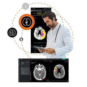
Instantly receive the information essential to your interpretation with our automated neurovascular emergency solution Optimize your management of stroke cases Standardize your emergency imaging workflow Boost confidence ...

... convenient possibilities of mediCAD® 3D Shoulder the individual implant components may be assembled and placed in the 3D model (CT scan of the patient) using the implant configurator. Additionally, the implants can be, ...
mediCAD Hectec

Once you open CT image data, mediCAD® 3D Spine performs an automatic segmentation of the 3D model. During this process, your recording is precisely analyzed and compared to the data repository of the software. ...
mediCAD Hectec

On CT images that have already undergone automatic segmentation, you can perform and document a number of conventional measurements automatically: - Scoliosis using Cobb’s method - Scoliosis using Ferguson’s method - ...
mediCAD Hectec

... decision support tool for assessing stroke signs on plain CT brain scans. Fully automates and standardizes the ASPECTS score and measures the volume of ischemic signs (ml) Assesses non-contrast CT ...

... advanced post-processing techniques in 2D and 3D, exclusive innovative technique for 3D & 4D navigation, including PET-CT and SPECT-CT support, and a complete integration with any PACS. It fully supports ...

Have you recently undergone a CT (computed tomography) or MRI (magnetic resonance imaging) examination? These medical imaging exams are generated by specialized radiology equipment, designed to create detailed images ...
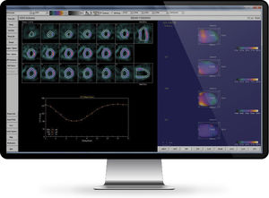
... nuclear cardiac review and reporting capabilities that enable physicians to quantify, review, and report SPECT, PET, Hybrid-CT, and myocardial blood flow and reserve studies. Both INVIA 4DM and Cedars-Sinai nuclear post-processing ...

Xelis™ Cardiac provides easy and powerful 2D and 3D display and analysis tools for evaluating coronary CT angiography. It delivers extensive volumetric studies with comprehensive image analysis, soft plaque analysis, ...

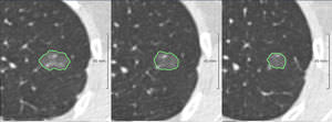
MeVis software solutions automatically detect abnormalities in computer tomography images - such as lung tumors or pulmonary embolism. Multi-slice computer tomography (MSCT) is state of the art for three-dimensional ...
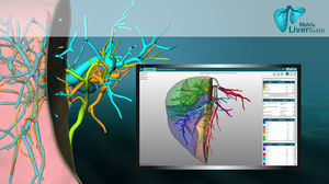
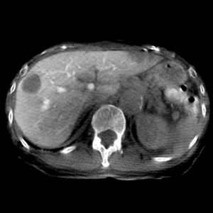
MeVis Liver Suite is a dedicated software solution to perform image analysis and visualization of hepatic and abdominal imaging studies derived from CT and MRI scanning devices. It is a powerful application ...
... The lung screening workstation from MeVis Veolity™ is the lung screening workstation for the efficient reading of chest CT images. When put to use in a high-throughput environment such as lung screening programs, ...
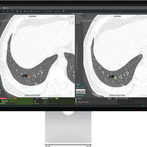
... according to 'Lung-RADS* ver 1.1' proposed by ACR as a lung nodule classification method. *Lung-RADS: Lung Cancer Screening CT Report And Data System *ACR: American College of Radiology Examine Follow-up data automatically By ...
... like USB, external hard drive or PACS. Image fusion enables the user to see the best information in each imaging modality (CT and MRI) combined, or the pre-operative cochlear parameter measurements with the post-operative ...
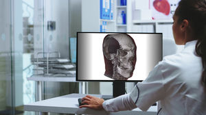
Complex software-assisted visualization methods enable the 3D visualization of image material from layer processes such as CT, MRI, or specific methods (e.g., OCT in ophthalmology). The previous 2-dimensional ...

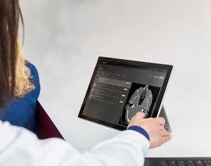
... internet browser and from all types of support ( digital tablet, computer, workstation) Diagnostic Web Viewer (MIP/MPR tools, PET-CT and PET-MR merge…) Clinical Web Viewer for clinicians for consultation reasons Sharing ...
... when the CT scan is routed. The code is printed to a selected network printer and can be presented to both patients and other clinicians. The client can be downloaded to run on most any machine on the same network ...
Novarad

Navigation Software Spine & Trauma streamlines your spine workflow Now, more than ever, it acts as a central data hub as it seamlessly switches between applications, a feature that is particularly important for a robotic ...
Brainlab
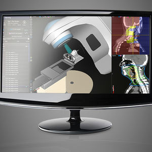
... clinical knowledge. Multi-Modality Image Registration Quick and easy to use rigid and deformable registration tools for CT, CBCT, MRI, PET, SPECT, AND RT images. Share data easily Sync with ARIA® OIS and Eclipse™ ...
Varian Oncology
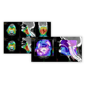
... systems, and automatically deforms previous treatment plan contours and previously delivered dose onto the new treatment planning CT image.
Please specify:
Help us improve:
remaining