
{{product.productLabel}} {{product.model}}
{{#if product.featureValues}}{{product.productPrice.formattedPrice}} {{#if product.productPrice.priceType === "PRICE_RANGE" }} - {{product.productPrice.formattedPriceMax}} {{/if}}
{{#each product.specData:i}}
{{name}}: {{value}}
{{#i!=(product.specData.length-1)}}
{{/end}}
{{/each}}
{{{product.idpText}}}
... detection systems and ancillary reagents to enable reliable RNA sequence detection.

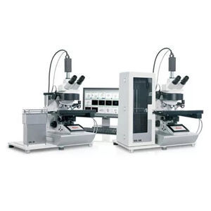
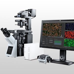
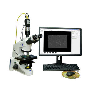
... demanding applications, the LAS X Widefield Systems offers high-speed acquisition, TIRF, Fura2, FRET SE, Screening, Ca2+ and ratio imaging, and peripheral triggering. The Ease-of-use Software Manual and automated ...
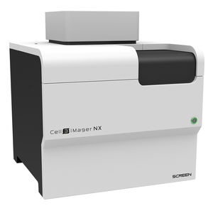
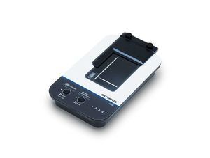
... another new addition to its range of high-quality cell analysis equipment. The Cell3iMager NX is an imaging system that captures and analyse 2D and 3D cultured cells ...
... streamlined and
automated
digital
cell morphology workflow, enabling laboratories to work smarter and perform better.
Developed to meet the needs of big labs with high-volume testin g requirements:
• ...
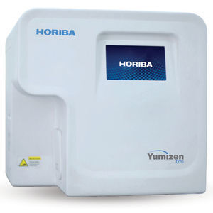
... and accurate results to meet patient's needs. The Yumizen D20 has 2 modules. Shonit: An AI application to analyze blood cell morphology which identifies and pre-classifies WBCs, RBCs and platelets in a peripheral blood smear. Shrava: ...
... The scanR modular microscope-based imaging platform provides fully automated image acquisition and data analysis of biological samples through deep-learning technology. Flexible, Modular Hardware The scanR screening ...
... data on cell health, cell count and confluency without labels using the automated CM30 incubation monitoring system. The CM30 enables non-invasive observation inside incubators, reduces ...
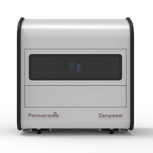
... br>3DHISTECH exhibited and demonstrated the Pannoramic Confocal system at AACR 2014. The system is a confocal digital slide scanner designed for optical sectioning and high-contrast imaging ...
... without full
system replacement.
Applications
- 3D cell imaging and analysis (organoids, spheroids)
- Cancer research and drug discovery
- Cell
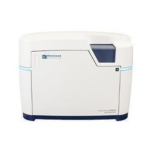
Molecular Devices
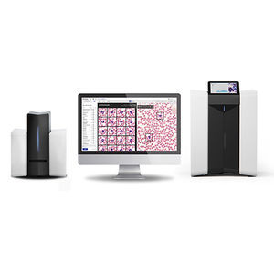
... Future of Digital Cell Morphology is Now 100X Closer Imaging tens of thousands of cells at a time, Scopio assists you in diagnosing hematological disorders by transforming the way ...
... into the automated processing of the EUROMicroblots. Due to the barcode scanner integrated in the instrument, reliable assignment of the EUROMicroblot plates is ensured. EUROLineScan, the proven software for automated ...
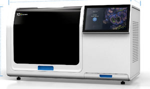
... A state-of-the-art single cell spatial imaging platform Seamlessly integrates transcript detection workflow, high-resolution imaging, decoding, and onboard data analysis. Built to deliver exceptional ...
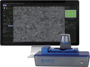
... Product Details The Live Cell Imaging System TOW-MCS-PRO is an excellent companion for cell culture. It can be used inside an incubator and supports phase contrast and fluorescence observation ...
Shanghai TOW Intelligent Technology

... another live- cell time-lapse microscopy system. The live- cell incubation system can be adapted to time-lapse microscopy to efficiently observe microscopic objects over time. Its main ...
Shanghai TOW Intelligent Technology
... during prolonged observations. Support for Live Cell Research This product is a critical component in building a live cell workstation, offering researchers the ideal tool for performing highly accurate cell ...
Shanghai TOW Intelligent Technology
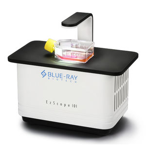
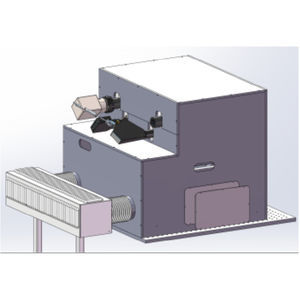
... EzScope 101 is a dedicated live cell imaging system that helps to streamline your research workflow with improved efficiency and productivity, no more hassles to remove cells from incubator ...
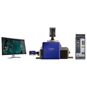
... Power Laser Engine (HLE) and new TIRF imaging modality, which exploits Borealis® illumination, B-TIRF (Borealis TIRF) for easy setup, more even illumination and thus more usable data across the field of view. Neuroscience Whether ...
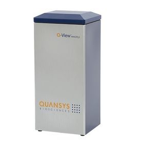
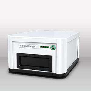
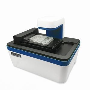
... The Q-View Imager LS is a high-quality, low-cost chemiluminescent imager that supports 96-well to 384-well plate-based chemiluminescent imaging. The Q-View Imager LS requires Q-View™ ...

... Satisfy everyone in your lab with an imager that does it all—and takes the complication out of getting reliable, fully analyzed data too! Meet the FluorChem™ R system. SENSITIVE See those tiny, hard to detect bands ...
ProteinSimple

... Automated high content image acquisition and analysis for drug discovery and cell biology Features The CELENA® X High Content Imaging System is an integrated imaging ...
Logos Biosystems

... F PRO is our latest cell-monitoring imager for continuous monitoring of bioprocesses. It is engineered to work with wave bags, single-use and stirred tank bioreactors and has been designed for cGMP compliance. Continuous ...
... or CEROS II analyzer at the main laboratory. This allows reduced costs while providing full services at all branches of a multi-site laboratory. The Remote Capture system is a standalone workstation for the capture ...
Hamilton Thorne
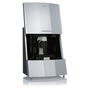
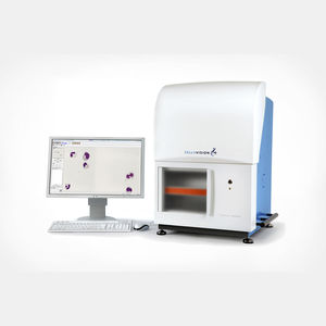
... physiological environment of cell cultures. Cell behavior and evaluation can be realized without staining. The system’s core technology uses high speed video imaging with a unique ...
Sony Biotechnology
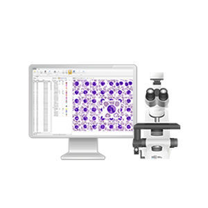
... large laboratories. Key features — Transfer of data from hematology analyzers or LIS — Processing of data based on the set rules and algorithms — Automatic decisions about the necessary actions with samples in ...
West Medica
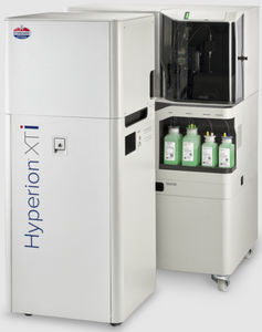
... Hyperion XTi™ Imaging System, the fastest and most reliable workflow for high-plex imaging. Discover the throughput and precision that is uniquely designed for translational researchers. Reasons ...
Fluidigm
... live cell imaging system is designed to get time-lapse images and make taking cell videos much easier. KEY FEATURES - Compact and compatible with a standard CO₂ incubator - Fully ...
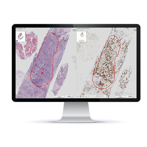
... A unique Brightfield imaging & analysis system for a variety of histopathology needs, including Quantitative IHC Scoring and Whole Slide Imaging of H&E/IHC samples. Through precise computer-assisted analysis ...
Applied Spectral Imaging
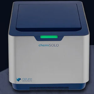
... ntroducing a new, personal chemiluminescent imager that delivers high-quality, quantitative chemiluminescent and visible protein gel imaging through a unique web-based control software Why Do Scientists Choose chemiSOLO? Smaller ...
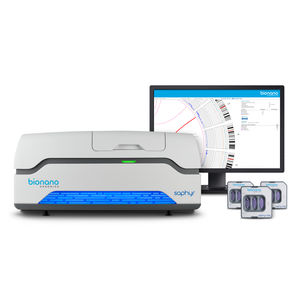
... to 500 base pair (bp) resolution and 5% variant allele frequency (VAF) with optical genome mapping (OGM) using the Saphyr system. Saphyr is the most powerful structural variant detection tool available, detecting genomic variants commonly ...
the best suppliers