
- Dental
- Dental practice
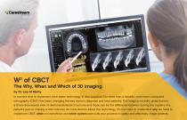
- Digital cephalometric X-ray system
- Carestream Dental
- Products
- Catalogs
- News & Trends
- Exhibitions
Digital cephalometric X-ray system CS 8100SC18x24 cm

Add to favorites
Compare this product
Characteristics
- Technology
- digital
- FOV (cm)
- 18x24 cm
Description
Overview
Carestream Dental's cephalometric module and software deliver high-accuracy cephalometric images and automated tracing with fast acquisition times. The solution is designed for orthodontic and oral surgery workflows and is offered across multiple product families to meet various clinical needs.
Key Benefits
Flexible Image Options
Supports all common projections and multiple image formats:
Fast Scans
Dedicated cephalometric sensor enables record-speed acquisition without sensor swapping between exams, reducing motion blur and exposure time.
Powerful Imaging Software
Intuitive imaging software enhances diagnostics and patient communication with advanced orthodontic filters that visualize hard and soft tissue with a single click.
Automatic Tracing
Carestream Dental automatic tracing capability:
Media Gallery (selected images / items)
Characteristics / Technical specifications
Carestream Dental's cephalometric module and software deliver high-accuracy cephalometric images and automated tracing with fast acquisition times. The solution is designed for orthodontic and oral surgery workflows and is offered across multiple product families to meet various clinical needs.
Key Benefits
- High accuracy and speed for cephalometric imaging
- Ultra-fast scanning technology to reduce motion blur and limit exposure time
- Automatic tracing to save clinician time and improve patient communication
- Multiple product-family options to suit different clinical environments
Flexible Image Options
Supports all common projections and multiple image formats:
- Image formats ranging from 26 cm x 24 cm to 18 cm x 18 cm (example image: 18 cm x 24 cm)
- Field collimation to restrict exposure to the area of interest
Fast Scans
Dedicated cephalometric sensor enables record-speed acquisition without sensor swapping between exams, reducing motion blur and exposure time.
Powerful Imaging Software
Intuitive imaging software enhances diagnostics and patient communication with advanced orthodontic filters that visualize hard and soft tissue with a single click.
Automatic Tracing
Carestream Dental automatic tracing capability:
- Fully traces images in approximately 10 seconds (example: 18 cm x 24 cm)
- Covers most common analyses including Ricketts, McNamara, Steiner and Tweed
Media Gallery (selected images / items)
- CS 9600 Scan Ceph Lateral
- CS 8100SC Pan Ceph Filter2
- CS 8100SC Evo Ceph Frontal AP
- CS 8100SC Evo Ceph Filter 2
- CS 8100 SC Evo Ceph Carpus
- CS 8100SC Ceph Submento Vertex
- CS 8100SC Ceph Frontal PA
- Ceph Video (product video / demo)
Characteristics / Technical specifications
- Supported image formats: from 26 cm x 24 cm down to 18 cm x 18 cm
- Dedicated cephalometric sensor (no sensor swapping between exams)
- Ultra-fast scan acquisition to minimize motion blur and reduce exposure time
- Field collimation to limit exposure to area of interest
- Automatic tracing time: approx. 10 seconds per tracing (example 18 cm x 24 cm)
- Automatic analyses supported: Ricketts, McNamara, Steiner, Tweed
- Advanced orthodontic image filters for enhanced hard- and soft-tissue visualization
Catalogs
Related Searches
- Analysis software
- Viewer software
- Radiology software
- Tablet computer software
- Flat panel sensor
- Control software
- Reporting software
- USB camera
- Diagnostic medical software
- Scheduling software
- Online software
- Automated software
- Dental software
- Design software
- Dental radiography system
- Treatment software
- Acquisition software
- Scan software
- Camera with LED light
- Data management software
*Prices are pre-tax. They exclude delivery charges and customs duties and do not include additional charges for installation or activation options. Prices are indicative only and may vary by country, with changes to the cost of raw materials and exchange rates.