- Primary care
- General practice
- Thorax model
- Erler-Zimmer
Thorax model MP1521for teaching
Add to favorites
Compare this product
fo_shop_gate_exact_title
Characteristics
- Area of the body
- thorax
- Procedure
- for teaching
Description
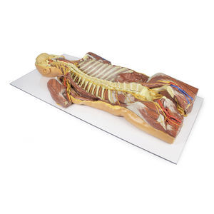
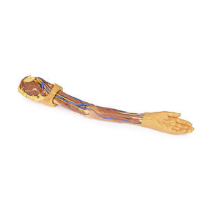
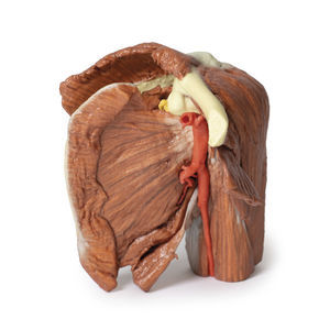
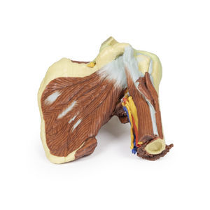
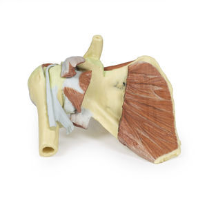
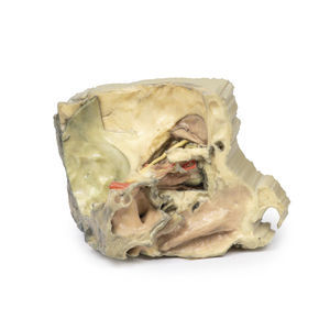
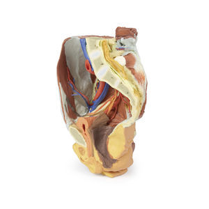
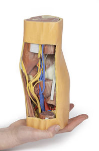
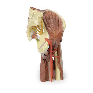
This 3D printed specimen preserves a dissection of the right thoracic wall, axilla, and the root of the neck. The specimen is cut just parasagittally and the visceral contents of the chest have been removed. Structures within the right chest wall are visible deep to the parietal pleura, including the ribs, muscles of the intercostal spaces and the origins of the neurovascular bundle in each intercostal space. The pectoralis major has been reflected medially towards the sectioned edge of the specimen to expose pectoralis minor which acts as a useful landmark as it divides the axillary artery into its three parts. The clavicle has had its middle 1/3 removed, but the subclavius muscle has been retained. The brachial plexus and many of its branches are seen almost in its entirety from the roots of C5-T1 to its termination as it exits the axilla to enter the arm.
Catalogs
Catalog 2021
360 Pages
3D Anatomy Series
9 Pages
Related Searches
- ERLER ZIMMER anatomical model
- ERLER ZIMMER training anatomical model
- ERLER ZIMMER teaching anatomical model
- Surgical anatomical model
- ERLER ZIMMER bone model
- ERLER ZIMMER flexible anatomical model
- ERLER ZIMMER skull model
- Denture model
- Plastic anatomy model
- Transparent anatomical model
- Dental anatomical model
- Oral anatomical model
- ERLER ZIMMER body anatomical model
- White anatomical model
- ERLER ZIMMER leg anatomical model
- ERLER ZIMMER spine anatomical model
- ERLER ZIMMER digestive system model
- ERLER ZIMMER pelvis model
- ERLER ZIMMER heart model
- ERLER ZIMMER nervous system model
*Prices are pre-tax. They exclude delivery charges and customs duties and do not include additional charges for installation or activation options. Prices are indicative only and may vary by country, with changes to the cost of raw materials and exchange rates.


