


- Secondary care
- Ophthalmology
- Retinal camera
- Optopol Technology
Non-mydriatic retinal camera FUNDOfundus imaging

Add to favorites
Compare this product
Characteristics
- Type of instrument
- non-mydriatic retinal camera
- Examination
- fundus imaging
Description
Overview
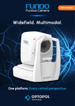
The FUNDO is a non-mydriatic automatic widefield fundus camera designed for efficient retinal diagnostics in clinical and outpatient settings. It offers true-color widefield imaging, multimodal acquisition and streamlined, automated workflows with AI-assisted prioritization to support fast, repeatable examinations.
Key imaging modalities
Field of view and widefield options
Standard single-capture angular field is 90° (60°) ±5%. Mosaic mode enables panoramic retinal coverage up to approximately 198° (132°) ±5% from the eye center using multiple fixation positions. A single 60° acquisition provides a broad overview for routine screening.
Automation and workflow features
Image quality and camera
Equipped with a 20.3 MP CMOS sensor, FUNDO delivers high-resolution images for detailed assessment of the retina, optic disc and retinal vasculature. Flash, gain and exposure are available in Auto and Manual modes with selectable intensity levels (High/Normal/Low).
Advanced fundus analysis tools
A suite of image processing and analysis tools supports clinical evaluation and follow-up:
Multimodal imaging details
AI support
Integrated AI algorithms automatically analyze acquired images to flag potential abnormalities and prioritize findings, aiding rapid image review and clinical decision-making.
Anterior segment & meibography
Anterior segment color imaging documents ocular surface, cornea, iris and pupil. Meibography provides non-invasive infrared imaging of Meibomian glands to support diagnosis of Meibomian Gland Dysfunction (MGD).
Connectivity & data management
Seamless integration with diagnostic software enables multi-station review, results management, DICOM compatibility, network connectivity and export to selected storage locations for an integrated diagnostic workflow.
Clinical use and design
The compact desktop design supports varied clinical configurations and flexible operator positioning (face-to-face, side-by-side or parallel). Non-mydriatic imaging enables comfortable exams without routine pupil dilation and supports imaging through small pupils.
Specifications / Technical data
The FUNDO is a non-mydriatic automatic widefield fundus camera designed for efficient retinal diagnostics in clinical and outpatient settings. It offers true-color widefield imaging, multimodal acquisition and streamlined, automated workflows with AI-assisted prioritization to support fast, repeatable examinations.
Key imaging modalities
- Color fundus photography (true-color)
- Infrared (IR) fundus imaging
- FAF – Green and Blue fundus autofluorescence
- Fluorescein angiography (FA)
- Indocyanine green angiography (ICGA)
- Anterior segment color photography
- Meibography (infrared imaging of Meibomian glands)
Field of view and widefield options
Standard single-capture angular field is 90° (60°) ±5%. Mosaic mode enables panoramic retinal coverage up to approximately 198° (132°) ±5% from the eye center using multiple fixation positions. A single 60° acquisition provides a broad overview for routine screening.
Automation and workflow features
- Automatic pupil detection, alignment, focus and exposure adjustment
- Automated image acquisition with optional voice guidance for consistent exams
- Internal OLED fixation target with adjustable shape, position and steady/blink modes; external fixation supported
- Alignment modes: Fully automatic, Automatic (assisted), Manual
Image quality and camera
Equipped with a 20.3 MP CMOS sensor, FUNDO delivers high-resolution images for detailed assessment of the retina, optic disc and retinal vasculature. Flash, gain and exposure are available in Auto and Manual modes with selectable intensity levels (High/Normal/Low).
Advanced fundus analysis tools
A suite of image processing and analysis tools supports clinical evaluation and follow-up:
- Single-eye and bilateral imaging
- Chronological comparison of fundus photographs (including x4 and x8 progression views)
- Linking of fundus images with OCT scans and visual field data for combined review
- Measurement tools: Area, Distance, Cup/Disc ratio (C/D)
- Image enhancement filters: Emboss+, Emboss−, Cobalt, Digital Cobalt, Digital Red-Free
Multimodal imaging details
- FAF Green for assessment of RPE and outer retina
- FAF Blue for complementary RPE and lipofuscin information
- Fluorescein angiography (FA) for retinal perfusion, leakage and ischemia analysis
- Indocyanine green angiography (ICGA) for choroidal circulation and deeper chorioretinal abnormalities
AI support
Integrated AI algorithms automatically analyze acquired images to flag potential abnormalities and prioritize findings, aiding rapid image review and clinical decision-making.
Anterior segment & meibography
Anterior segment color imaging documents ocular surface, cornea, iris and pupil. Meibography provides non-invasive infrared imaging of Meibomian glands to support diagnosis of Meibomian Gland Dysfunction (MGD).
Connectivity & data management
Seamless integration with diagnostic software enables multi-station review, results management, DICOM compatibility, network connectivity and export to selected storage locations for an integrated diagnostic workflow.
Clinical use and design
The compact desktop design supports varied clinical configurations and flexible operator positioning (face-to-face, side-by-side or parallel). Non-mydriatic imaging enables comfortable exams without routine pupil dilation and supports imaging through small pupils.
Specifications / Technical data
- Type: Non-mydriatic automatic fundus camera
- Photograph types: Color Fundus/Anterior, IR Fundus/Anterior, FAF – Green*, FAF – Blue*, FA*, ICGA*
- Angular field of view (standard pupil): 90° (60°) ± 5%
- Angular field of view (small pupil): 67° (45°) ± 5%
- Mosaic field of view (standard pupil): 198° (132°) ± 5%*
- Minimum pupil size: 3.3 mm (small pupil)
- Camera: 20.3 MP CMOS
- Flash adjustment / gain / exposure: Auto, Manual
- Intensity levels: High, Normal, Low
- Focus adjustment range: -25 D to +25 D
- Operating distance: 31 mm
- Alignment method: Fully automatic, Automatic, Manual
- Internal fixation target: OLED display with adjustable target shape, position, steady/blinking light mode and blink rate
- External fixation target: Yes
- Dimensions (L×W×H): 490 × 350 × 530 mm
- Weight: 19 kg
- Power supply: 100–240 V, 50/60 Hz
- Power consumption: 90–110 VA
- Optional features: Certain modes (marked with *) are optional and selectable during ordering (e.g., some FAF/angiography and mosaic capabilities)
Other Optopol Technology products
SD OCT | Fundus Camera Combo


*Prices are pre-tax. They exclude delivery charges and customs duties and do not include additional charges for installation or activation options. Prices are indicative only and may vary by country, with changes to the cost of raw materials and exchange rates.
About MedicalExpo
MedicalExpo is a marketplace that offers B2B players a complete experience, from product sourcing to purchase. MedicalExpo is an entity of VirtualExpo Group.
Are you a buyer?
Are you a supplier?
© 2026 All rights reserved - Terms - Privacy policy - General Terms - Manage cookies - 鄂ICP备2026053537号-3 -