


{{product.productLabel}} {{product.model}}
{{#if product.featureValues}}{{product.productPrice.formattedPrice}} {{#if product.productPrice.priceType === "PRICE_RANGE" }} - {{product.productPrice.formattedPriceMax}} {{/if}}
{{#each product.specData:i}}
{{name}}: {{value}}
{{#i!=(product.specData.length-1)}}
{{/end}}
{{/each}}
{{{product.idpText}}}
{{product.productLabel}} {{product.model}}
{{#if product.featureValues}}{{product.productPrice.formattedPrice}} {{#if product.productPrice.priceType === "PRICE_RANGE" }} - {{product.productPrice.formattedPriceMax}} {{/if}}
{{#each product.specData:i}}
{{name}}: {{value}}
{{#i!=(product.specData.length-1)}}
{{/end}}
{{/each}}
{{{product.idpText}}}
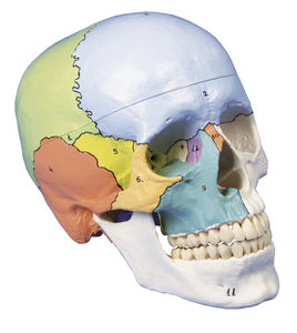

Length: 18 cm
Width: 19 cm
Height: 12 cm
... Skull model like ref.no. 4500, additionally with didactical painting of individual bones on one side of the skull. The bones are numbered referring to the included nomenclature. ...
Erler-Zimmer

... This skull model is an actual cast of a real human specimen and shows all anatomical structures in highest detail. It is made for students in anatomy, medicine, surgery, otolaryngology, ophthalmology and dentistry. The Skull is intricately ...
Erler-Zimmer

Length: 15 cm
Width: 15 cm
Height: 30 cm
... This life size head model is dissected along the sagittal plane into 2 halves. Details of the oronasal cavity and larynx as well as musculature of the pharynx are exceptionally well represented. Mounted on base with stand. ...
Erler-Zimmer

Length: 50 cm
Width: 20 cm
Height: 41 cm
... ventricles, and choroid plexus. The right side retains skin and fascia, false-coloured to highlight facial and neck angiosomes. The left side reveals facial expression and mastication muscles, and infratemporal structures ...
Erler-Zimmer

... This unique 3D model is derived from dural skull (Print #8) data, obtained via CT imaging and segmentation of internal spaces. Parts of the skull have been retained, while sections or windows have been removed to expose the paranasal ...
Erler-Zimmer

... This 3D print provides a detailed view of head and neck anatomy, showing different structures depending on the perspective. Lateral Face and Parotid Region A window exposes the parotid region, with the pinna intact ...
Erler-Zimmer

... infratemporal fossa and surrounding neck structures. Mandible and Infratemporal Fossa The ramus, coronoid process, and mandibular head have been removed to expose the deep infratemporal fossa. The pterygoid muscles are removed to ...
Erler-Zimmer

... provides a detailed exploration of the head and visceral column of the neck, showcasing muscles, nerves, arteries, veins, and cranial structures. Facial Anatomy The right side of the face is dissected ...
Erler-Zimmer

... nasociliary nerve, ophthalmic artery, and superior ophthalmic vein within the intraconal space. Facial Anatomy The face has been dissected to show facial muscles around the orbit on the right, while ...
Erler-Zimmer
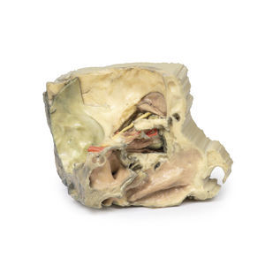
... path as it enters the posterior ethmoidal foramen. Additional Anatomical Features Other key structures visible in this model include the frontal nerve (FN), sphenoid sinus (SS), pituitary gland (PG), and the frontal sinus mucosal ...
Erler-Zimmer