
{{product.productLabel}} {{product.model}}
{{#if product.featureValues}}{{product.productPrice.formattedPrice}} {{#if product.productPrice.priceType === "PRICE_RANGE" }} - {{product.productPrice.formattedPriceMax}} {{/if}}
{{#each product.specData:i}}
{{name}}: {{value}}
{{#i!=(product.specData.length-1)}}
{{/end}}
{{/each}}
{{{product.idpText}}}

... transsubclavian, transcarotid, transapical or direct aortic approach is most suitable. The software has an intuitive workflow assistant which acts as a guide through the software making it intuitive and ...
Pie Medical Imaging

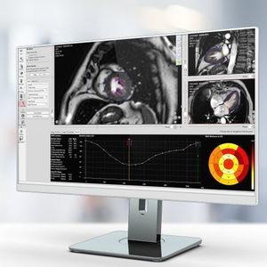
Furthermore, the 3D reconstruction enables assessment of severity and percentage of stenosis. Functional lesion information -Pressure drops in coronary artery -vFFR value Anatomical lesion information -Lesion diameter -Percentage ...
Pie Medical Imaging
... displacement Key product features Single click vessel border detection Give anatomical designation for direct calculation of clinical results Aliasing and offset correction Analyze up to four phase contrast image sets
Pie Medical Imaging
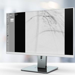
StentEnhancer features enhanced stent visualization. The application automatically enhances the image region where the stent is deployed.
Pie Medical Imaging

... analysis The Wall Shear Stress Analysis and Pressure Difference Analysis are not 510(k) cleared and therefore not meant for clinical decision making. Key product features Intracardiac analysis with automated valve ...
Pie Medical Imaging
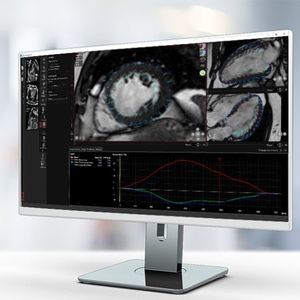
The endocardial and epicardial wall of the ventricle can be automatically segmented on short axis images. Long axis images can also be used for segmentation. Ejection fraction, End Diastolic (ED), and End Systolic (ES) volumes are accurately ...
Pie Medical Imaging

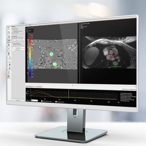
Caas MR Tissue Characterization includes three workflows: Viability, First-Pass Perfusion, and Tissue Mapping. Differentiate between viable and non-viable tissue using regional infarct classification based on delayed-enhanced MR images. ...
Pie Medical Imaging
... Tracking Detect ventricular deformation patterns and functional abnormalities Global longitudinal strain (GLS) with high clinical value in: Systolic dysfunction (HFrEF, Cardiomyopathy) Dilated and Hypertrophic Cardiomyopathy Ischemic ...
Pie Medical Imaging

... Multiple custom catalogs may be created, for example based on your center’s standard operating procedures (SOPs) for a specific clinical indication or patient group. Myocardial strain analysis Our validated myocardial ...
Pie Medical Imaging
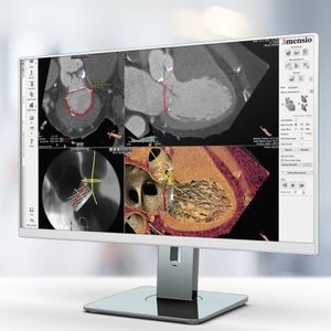
The Mitral Valve is a complex 3D structure. Mitral regurgitation can be treated by replacing the native valve (TMVR) or by repairing the native valve (TMVr). Planning for both types of procedures can be done using the 3mensio Mitral Valve ...
Pie Medical Imaging
Please specify:
Help us improve:
remaining