Analysis software visualizationmedicalclinical
Add to favorites
Compare this product
fo_shop_gate_exact_title
Characteristics
- Function
- analysis, visualization
- Applications
- medical, clinical, for MRI, cardiology
- Area of the body
- heart
- Deployment mode
- for tablet PC
Description
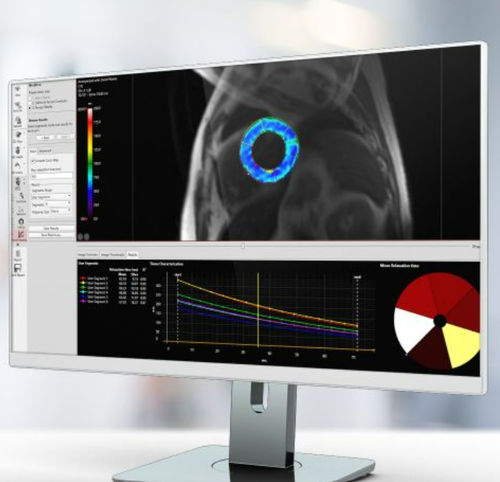
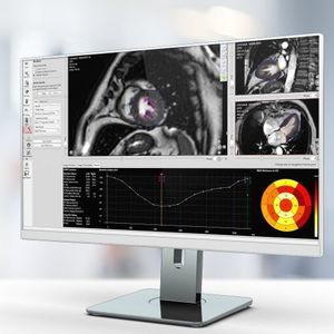
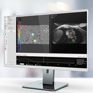
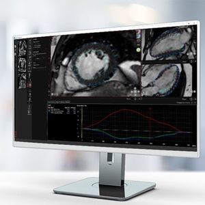
Caas MR Tissue Characterization includes three workflows: Viability, First-Pass Perfusion, and Tissue Mapping. Differentiate between viable and non-viable tissue using regional infarct classification based on delayed-enhanced MR images. Assess myocardial edema based on T2-weighted images. By combining segmental infarct and edema areas, salvageable areas in the area at risk can be identified. Analyze rest and stress perfusion image sets side by side in the first-pass perfusion workflow to determine myocardial perfusion. Tissue mapping by discriminating between T1 and T2 tissue contrast is a unique strength of MRI. It allows the analysis of T1, T2, and T2* relaxation values. Relaxation values are translated into a color map for easy visualization of affected tissue. Visualization of T1 relaxation values is supported for look-locker and modified look-locker sequences.
Key Results:
Infarct volume and transmurality
Salvageable area index
Time-Intensity curve parameters
Myocardial Perfusion Reserve Index (MPRi) derived by comparing rest and stress
T1, T2, T2* relaxation values
Parametric color maps
Key product features
Automatic infarct detection
Breathing motion correction
All result types are supported by the AHA 17-segment model
Catalogs
No catalogs are available for this product.
See all of Pie Medical Imaging‘s catalogsRelated Searches
- Analysis medical software
- Radiology software
- Viewer software
- Tablet PC software
- Windows medical software
- Reporting software
- Scheduling software
- Diagnostic medical software
- Automated software
- Hospital software
- Treatment software
- Tracking software
- Software module
- Measurement software
- Import software
- On-premise software
- CT software
- Server software
- Cardiac software
- Image analysis software
*Prices are pre-tax. They exclude delivery charges and customs duties and do not include additional charges for installation or activation options. Prices are indicative only and may vary by country, with changes to the cost of raw materials and exchange rates.


