
{{product.productLabel}} {{product.model}}
{{#if product.featureValues}}{{product.productPrice.formattedPrice}} {{#if product.productPrice.priceType === "PRICE_RANGE" }} - {{product.productPrice.formattedPriceMax}} {{/if}}
{{#each product.specData:i}}
{{name}}: {{value}}
{{#i!=(product.specData.length-1)}}
{{/end}}
{{/each}}
{{{product.idpText}}}
... Overview
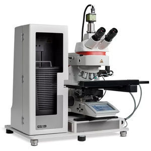
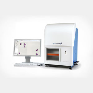
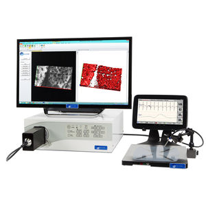
The CytoInsight GSL automated image analysis and case management
system is a comprehensive Cytogenomics solution that improves quality and turnaround time from acquisition to analysis while reducing hands-on time at ...
... Product overview
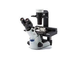
The CKX53 inverted microscope is designed for
cell and tissue culture workflows, simplifying live-
cell observation,
cell sampling and handling, image capture and fluorescence ...
Evident - Olympus Scientific Solutions
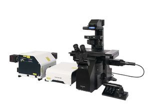
... super-resolution microscope system. Engineered for prolonged cell viability during time-lapse experiments, the IX85 SpinSR integrates fast image acquisition and advanced reconstruction algorithms to deliver XY resolution ...
Evident - Olympus Scientific Solutions
... The scanR modular microscope-based imaging platform provides fully automated image acquisition and data analysis of biological samples through deep-learning technology. Flexible, Modular Hardware The scanR screening station combines ...
Evident - Olympus Scientific Solutions

... The LAS X Widefield Systems are ideal for applications in fluorescence microscopy and image analysis including live cell time-lapse experiments, multi-positioning, z-stacking and deconvolution. The versatile, fully ...
... the process of performing blood and body fluid differentials. The system leverages high-speed robotics and digital imaging to automatically locate and capture high-quality images of cells. When implemented ...
... An in-vitro diagnostic device designed to automate manual microscopy in a diagnostic laboratory. It uses robotics and AI to digitize Blood and Urine samples to enable AI aided remote review. It generates a pre-classified report of blood ...
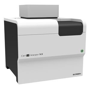
... another new addition to its range of high-quality cell analysis equipment. The Cell3iMager NX is an imaging system that captures and analyse 2D and 3D cultured cells ...
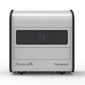
... demonstrated the Pannoramic Confocal system at AACR 2014. The system is a confocal digital slide scanner designed for optical sectioning and high-contrast imaging of histology and fluorescence samples, ...
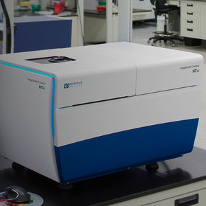
... The ImageXpress® Confocal HT.ai High-Content Imaging System utilizes a seven-channel laser light source with eight imaging channels to enable highly multiplexed assays while maintaining ...
Molecular Devices
... A state-of-the-art single cell spatial imaging platform Seamlessly integrates transcript detection workflow, high-resolution imaging, decoding, and onboard data analysis. Built to deliver exceptional ...
... Product Details The Live Cell Imaging System TOW-MCS-PRO is an excellent companion for cell culture. It can be used inside an incubator and supports phase contrast and fluorescence observation ...
Shanghai TOW Intelligent Technology

... another live- cell time-lapse microscopy system. The live- cell incubation system can be adapted to time-lapse microscopy to efficiently observe microscopic objects over time. Its main ...
Shanghai TOW Intelligent Technology
... during prolonged observations. Support for Live Cell Research This product is a critical component in building a live cell workstation, offering researchers the ideal tool for performing highly accurate cell ...
Shanghai TOW Intelligent Technology
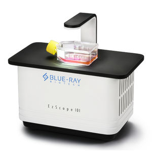
... EzScope 101 is a dedicated live cell imaging system that helps to streamline your research workflow with improved efficiency and productivity, no more hassles to remove cells from incubator ...
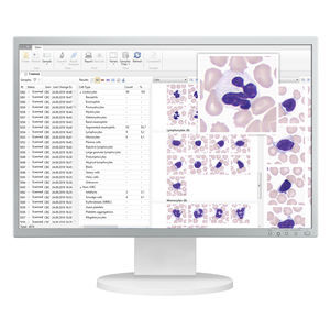
... Automatic animal blood cells analysis Identification and pre-classification of leucocytes — Basophils — Eosinophils — Promyelocytes — Myelocytes — Band neutrophils — Segmented neutrophils — Lymphocytes — Monocytes — ...
West Medica
... Power Laser Engine (HLE) and new TIRF imaging modality, which exploits Borealis® illumination, B-TIRF (Borealis TIRF) for easy setup, more even illumination and thus more usable data across the field of view. Neuroscience Whether ...
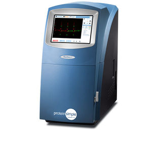
... in FluorChem imagers ensure that you'll get the best image quality and the resolution you need to do accurate analysis, and differentiate between bands that are close together. The FluorChem M and R also enable multiplex fluorescent imaging, ...
ProteinSimple

... discovery and cell biology Features The CELENA® X High Content Imaging System is an integrated imaging system designed for rapid, high content image acquisition ...
Logos Biosystems

... F PRO is our latest cell-monitoring imager for continuous monitoring of bioprocesses. It is engineered to work with wave bags, single-use and stirred tank bioreactors and has been designed for cGMP compliance. Continuous ...
... station lets satellite laboratories process samples and save video for analysis on an IVOS® II or CEROS II analyzer at the main laboratory. This allows reduced costs while providing full services at all ...
Hamilton Thorne
... physiological environment of cell cultures. Cell behavior and evaluation can be realized without staining. The system’s core technology uses high speed video imaging with a unique ...
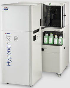
... Hyperion XTi™ Imaging System, the fastest and most reliable workflow for high-plex imaging. Discover the throughput and precision that is uniquely designed for translational researchers. Reasons ...
Fluidigm
... live cell imaging system is designed to get time-lapse images and make taking cell videos much easier. KEY FEATURES - Compact and compatible with a standard CO₂ incubator - Fully ...
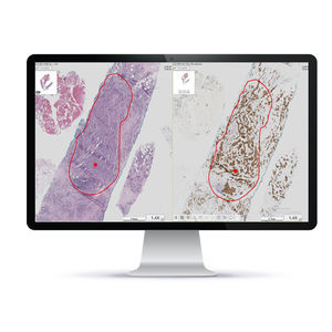
... A unique Brightfield imaging & analysis system for a variety of histopathology needs, including Quantitative IHC Scoring and Whole Slide Imaging of H&E/IHC samples. Through precise computer-assisted analysis ...
Applied Spectral Imaging
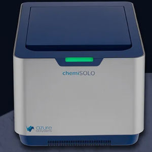
... ntroducing a new, personal chemiluminescent imager that delivers high-quality, quantitative chemiluminescent and visible protein gel imaging through a unique web-based control software Why Do Scientists Choose chemiSOLO? Smaller ...
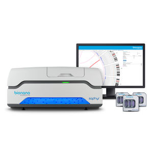
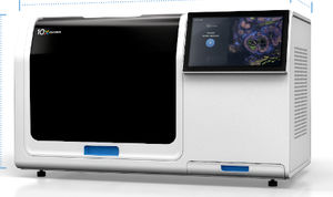
... to 500 base pair (bp) resolution and 5% variant allele frequency (VAF) with optical genome mapping (OGM) using the Saphyr system. Saphyr is the most powerful structural variant detection tool available, detecting genomic variants commonly ...
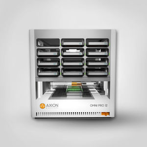
... fluorescence live- cell imaging platform for drug discovery and development. Featuring integrated robotics, automated plate handling, and powerful software with foolproof data storage, the Omni Pro 12 enables continuous, ...
Axion BioSystems

... Results Ultimate linearity for precise protein quantification over the full dynamic range. Multispectral Imaging Ultra-low noise imaging thanks to a dual camera amplifier architecture. Custom Made V.070 Lens Fusion ...
Vilber GmbH
... MRS CellLIVE is a powerful, handheld fluorescence confocal endomicroscope imaging system that is designed specifically for in vivo research of a variety of animal models in a broad range of studies and ...
the best suppliers