- Laboratory
- Laboratory medicine
- Automatic cell imaging system
- Caliber Imaging & Diagnostics

- Products
- Catalogs
- News & Trends
- Exhibitions
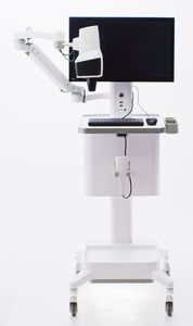
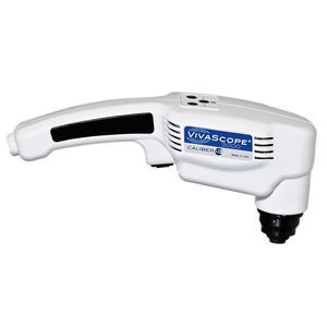
Automatic cell imaging system VivaScope® 2500for scientific researchpreclinicaltissue
Add to favorites
Compare this product
Characteristics
- Operation
- automatic
- Applications
- for scientific research, preclinical, tissue
- Cell type
- skin epithelial cells
- Observation technique
- fluorescence, confocal, phase contrast, infrared
- Other characteristics
- high-resolution
Description
The VIVASCOPE 2500 is a confocal microscope specially designed for imaging fresh, needle aspirated, or fixed specimens in reflectance (phase contrast) mode or in fluorescence mode for specimens stained with fluorochromes. Specimens can be examined in near real-time without time consuming processing procedures. The VIVASCOPE 2500 is used by physicians and other licensed healthcare professionals to view enlarged images of specimens during pathological examinations.
Features
Two lasers:
488nm (blue)
785 nm (infrared)
Filter sets for fluorescent dyes including:
Acridine orange
Fluorescein
Indocyanine green
Offers high-resolution photo of specimen
Correlates to confocal image for navigation
Simplifies selection of imaged area
SINGLE IMAGE
The VIVASCOPE 2500 generates single images of tissue that are parallel to the tissue surface or on the “horizontal plane” .
The image depth, or position within the specimen, is changed by moving the objective lens up, down or laterally, relative to the specimen surface. -
STACK
A series of horizontal images captured in “Z-depth”, or from the surface of the specimen to a preconfigured depth. The image plane is incrementally changed by moving the objective lens of the VIVASCOPE 2500 and images are captured at consecutively deeper depths within the tissue.
MOSAIC
An array of images taken on a single horizontal plane creates a mosaic. The VIVASCOPE 2500 scans the objective lens laterally over the tissue while capturing a series of images and displaying them as a composite image covering an area up to 20 x 20mm.
Technical Data
Optical Section Thickness - <4μm
Single Field of View Size - 550μm x 550μm
Catalogs
Other Caliber Imaging & Diagnostics products
CLINICAL IMAGING
Related Searches
- Microscopy
- Compound microscope
- Laboratory microscope
- Tabletop microscope
- Viewer software
- Laboratory software
- Online software
- Scan software
- Zoom microscope
- Research microscope
- Sharing software
- Automated cell imaging system
- Cell imager
- Interpretation software
- IR microscope
- Laboratory cell imaging system
- Confocal microscope
- Laser microscope
- Fluorescence cell imaging system
- Diagnostic cell imaging system
*Prices are pre-tax. They exclude delivery charges and customs duties and do not include additional charges for installation or activation options. Prices are indicative only and may vary by country, with changes to the cost of raw materials and exchange rates.


