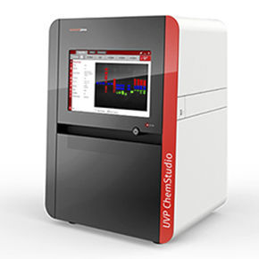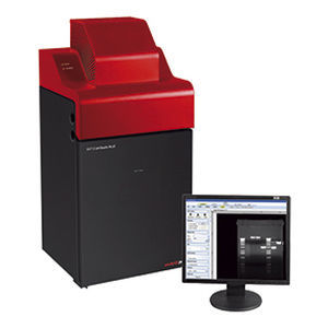
Fluorescence preclinical imaging system iBox® Explorer²™for small animalsfor in-vivo imaging
Add to favorites
Compare this product
Characteristics
- System type
- fluorescence
- Application
- for small animals, for in-vivo imaging
Description
Macro to Micro Fluorescent In Vivo Small Animal Imaging
Easy transition from the macroscopic to the microscope scale
Visualize micro injection of cancer cells in vivo
Ability to image organs and cells subcutaneously and within the body cavity of living mice
Optical configurations are parcentered and parfocal
Product details
Now researchers can take detection of fluorescent markers in small animals to a new level with the iBox Explorer² Imaging Microscope! The iBox Explorer² is unique in its ability to view macro to micro in the whole animal to individual cell, subcutaneously and within the body cavity of mice. The upright optics provide an ultra long working distance and high numerical aperture (NA) for detailed fluorescent in vivo imaging.
The UVP iBox Explorer² easily detects GFP/RFP and other fluorescent markers in small animals. The capabilities of this in vivo small animal imaging system enables research studies in whole animal down to individual cells.
Focusing on Fluorescent Cancer Cell Detection
In research performed, HT1080 fluorescent cancer cells were injected into the epigastrica cranialis. Immediately after the injection, the fluorescence indicates the cells escaped around the injection sites. In the zoomed in views, a cluster of cancer cells can be observed in the blood stream.
System Capabilities
Capture images with a superior cooled CCD camera and optics that are optimized for VIS - NIR imaging
View the whole animal to the single cell level via the motorized optics
Catalogs
No catalogs are available for this product.
See all of UVP‘s catalogs*Prices are pre-tax. They exclude delivery charges and customs duties and do not include additional charges for installation or activation options. Prices are indicative only and may vary by country, with changes to the cost of raw materials and exchange rates.


