
{{product.productLabel}} {{product.model}}
{{#if product.featureValues}}{{product.productPrice.formattedPrice}} {{#if product.productPrice.priceType === "PRICE_RANGE" }} - {{product.productPrice.formattedPriceMax}} {{/if}}
{{#each product.specData:i}}
{{name}}: {{value}}
{{#i!=(product.specData.length-1)}}
{{/end}}
{{/each}}
{{{product.idpText}}}
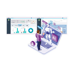
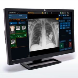
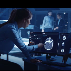
Tools to power up your business. Di.V.A. is an advanced digital assistance platform that allows constant monitoring of extraoral radiographic equipment. Detailed statistics on use, performance and maintenance give professionals full ...
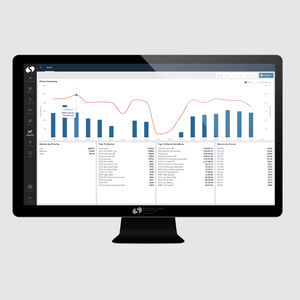
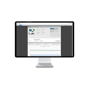
Maximize efficiency Spacelabs SafeNSound provides tools for efficient device management, streamlined communications between caregivers, and simplified admissions. Optimize throughput by decreasing device utilization Decrease communications ...
Spacelabs Healthcare
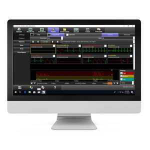
... carries vital information to clinicians across services and sites. ICS can also deliver clinical information to your desktop PC and mobile devices. Working seamlessly with your EMR systems, ICS supplies both numeric ...
Spacelabs Healthcare
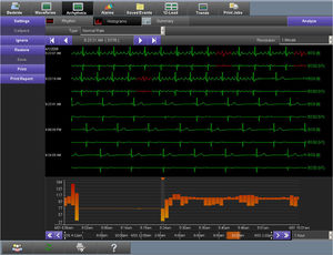
... Consolidated patient view Access waveform data, alarms, vital sign trends, and 12-lead ECGs for a specific time period from any PC on the network. Complete alarm history With a simple touch of a button, you can see ...
Spacelabs Healthcare

The Planmeca Romexis® Cephalometric Analysis software module is the ultimate tool for performing cephalometric analyses, surgical planning and treatment follow-ups in 2D. The software provides benefits ...
Planmeca

Planmeca Romexis® Ortho Simulator is an intuitive software module for orthodontic simulation. It allows using Planmeca Emerald® S intraoral scans to reveal the true potential of a patient’s smile in a matter of minutes. ...
Planmeca
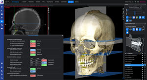
The Planmeca Romexis® 3D Cephalometry software module is the leading-edge tool for performing orthodontic analysis using CBCT images. The intuitive and easy tracing of anatomical landmarks as well as the clear visual ...
Planmeca

... used for every image and that the software can be used intuitively and logically. We attach special importance to perfectly supporting the efficiency of DÜRR MEDICAL systems, be it CR or DR Why DÜRR MEDICAL software DÜRR ...

W20s is a software that records the signals obtained through the SIBELMED spirometer, so that they can later be analyzed and saved in a simple and intuitive way. It is compatible with the DATOSPIR range, and can operate ...

SIBELMED W50 is a software for viewing, storing, transferring, analyzing and introducing audiometric tests, compatible with the range of SIBELMED audiometers. Audiometric charts With the test results Different ...

BitmedLab is a simple and intuitive software for viewing and reviewing sleep signals, and for the analysis of respiratory events. BitmedLab complies with the recommendations of the AASM, and is compatible with the ...

... and production analysis. Easy Reporting and Export Generate simple and clear reports of weighing results. Export data to PC in various formats (XML, CSV, XLSX or PDF) or print on a network printer.

... , obtained from high-throughput Aperio scanners, and retain precision quality with Aperio eSlide Manager image management software with WebViewer DX interface. Designed to optimize digital pathology processes for pathologists, ...
DR4 is a unique vascular reporting software package for use in conjunction with the Dopplex Ability, MD2 and MD200 Doppler units. It enables automated ABIs and Doppler vascular studies to be undertaken and saved in a ...

From seamless and precise data delivery through an accessible user interface to intuitive controls, our hemodynamic and electrophysiology recording systems are proven and time-tested. They help you treat the most difficult cardiac conditions ...
GE Healthcare
the all-in-one software platform for 2D and 3D imaging, is DATA PROTECTION certified and IHE compliant with DICOM networks. iRYS is a tool that provides dentists with an array of functions that lets them view, process ...
MUSICA™ Flex for unprecedented flexibility to match the intelligent image processing to the radiologist’s tastes… MUSICA™ Analytics transforms data into Quality and Performance insights. Digital Tomosynthesis(*) for multi-slice image ...
Artificial Intelligence is no longer an abstract promise. The Eclipse engine that powers our ImageView software puts AI into indisputable action — driving concrete, measurable results through Imaging Intelligence, Workflow ...
Carestream

Get a complete overview and centralized control of your point-of-care testing setup Grant device access to qualified operators Consolidate devices’ status on dashboard Validate device performance with peers In your daily work ...

... we can optimize your analyzer uptime and enhance your user experience. Remote Support Remote troubleshooting Remote software update* No patient or patient ID-related data are visible or accessible at any time. For ...
Inspect QC results daily and spot issues early Meet compliance requirements with monthly QC reports Reduce costs with a cloud-based QC management solution Streamline your QC data management workflow Strong Quality Control (QC) ...

The comprehensive EyeSuite IOL includes a wide range of IOL calculation formulae for any patient including toric and post-refractive cases. It is fully integrated into both the Eyestar 900 and Lenstar 900 biometers. Thus, it simplifies ...

... with the best computer-aided software analysis tools Multi-disciplinary collaboration and online continued medical education frameworks We offer fully-fledged software applications* and OEM-facing ...
VAREX Imaging

... through e-mail. All of the relevant tooth shade information and patient photos can be sent conveniently from a smartphone or tablet. VITA mobileAssist + enables the efficient communication of tooth shade information ...

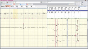
NeuroWorks EEG software simplifies the process of collecting, monitoring, trending and managing EEG testing data, allowing care providers to save time and focus on delivering the best care. As the EEG platform trusted ...
Natus Medical
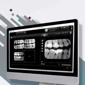
Discover a new generation of dental imaging software with CS Imaging version 8—the platform that provides one-stop access to all your 2D images, 3D images and CAD/CAM data—to manage your digital workflow more effectively. Save ...
Carestream Dental
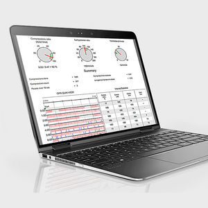
... lives into targeted improvements — CODE-STAT data review software and service lets you easily understand team performance immediately after response. Better data means better CPR CODE-STAT software ...
Bringing an additional layer of intelligence to our Collaborative imaging offering, Automation Platform is an AI-based, zero-click solution that uses deep learning technology to streamline your workflow for fast, actionable results every ...
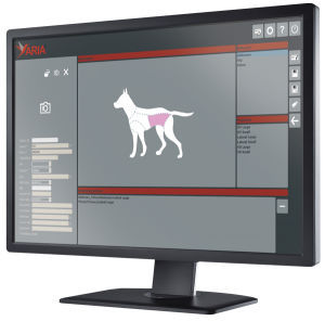
Through an extremely intuitive user interface, ARIA enables fast access to records and x-ray exams. You can search using multiple parameters including owner, name, gender, species and more. In just three steps you can select the study ...

Instructor-led virtual learning that delivers realistic clinical education, regardless of learner location Meet Maestro Evolve Deliver powerful simulation training with Maestro Evolve, an interactive virtual learning platform with ...

... tools and sophisticated archiving functionality. BRAIN QUICK EEG software is adaptable to any size facility, from large hospitals to private practice and research centers. Software Key Features: SQL ...
NEUROWERK software forms the basis for successful work with our NEUROWERK EMG and EEG systems. Thanks to its powerful SQL database, it provides easy access to all functions of our devices. Work more efficiently With ...

... SleepRT™ is the only clinical software package available worldwide which includes automatic calculation of REM Sleep Behavior Disorder. Both tonic and phasic EMG activity during REM is automatically calculated by the ...

... DICOM & HL7 COMPLIANT XC's software standards allow you to automatically send images to PACS. it can be integrated with patient management systems including RIS, HIS, and EMR. DICOM & HL7 COMPLIANT XC's software ...
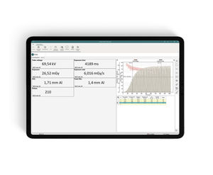
The world-leading traceability software for X-ray QA, testing, and reporting from RTI has been upgraded! Ocean Next™ software is the interface, via your RTI meter or probe, through which you directly ...
Please specify:
Help us improve:
remaining