
- Company
- Products
- Catalogs
- News & Trends
- Exhibitions
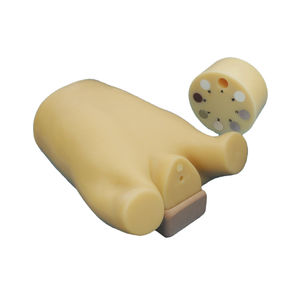
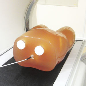
Nuclear imaging test phantom PH-63thorax
Add to favorites
Compare this product
Characteristics
- Type of calibration
- for nuclear imaging
- Area of the body
- thorax
Description
Examination of myocardial density through SPECT imaging
>Verification of myocardial imaging with the use of various RI solution densities
>Ability to capture defects of the myocardial region
>Can reproduce image variations of the heart by injecting RI solutions in the liver, kidney and lungs
Examination of RI solution density for simulated tumors
>The simulated tumors can be inserted into lung, liver andbreast
>Tumors can be filled with FDG/RI solution into the spheresfor evaluation of density, size and placement
Training skills / Applications - PET/SPECT
Quality management of NM equipment
Myocardial density with SPECT imaging
RI solution density for tumor imaging
Case / Pathology - >Anatomy
Liver
Lung (right/left)
Kidney (right/left)
Hot spots (liver, lungs and breast)
* Hot spot for PET can be set in liver, lungs and breast.
Heart
- Anatomical type:
right ventricle, left ventricle
and myocardium
- Geometric type:
left ventricle and myocardium
Set includes - 1 thorax body
2 lungs (left and right)
4 hearts
1 liver
2 kidneys
1 rib cage and spine
2 breasts
3 hot spots
1 base
several plastic pins
6 supporting bars
4 flat bar rings for base
5 tubes
1 syringe
several nuts and bolts
1 water tank
manual
1S6451S1
Size (approx.) - W44×D29×H71 cm
Weight (approx.) - 約21 kg (ファントム本体)、 約40 kg (満水時)
Materials - Soft tissue: transparent polyurethane
Lungs: materials with density 0.4 g/cm3
Bone materials: Calcium infused material to provide proper
attenuation with use of RI solutions
***********
HU
Bone: 370HU
Lung: -900HU
Organ shell material: 100HU, and 1.16g/cm3 in density
Catalogs
Radiology Phantoms
52 Pages
Related Searches
- Calibration phantom
- Tomography test phantom
- Radiography test phantom
- CT scan test phantom
- General purpose test phantom
- Ultrasound imaging test phantom
- MRI test phantom
- Torso test phantom
- Head test phantom
- Radiation therapy calibration phantom
- Breast test phantom
- Abdomen test phantom
- Mammography test phantom
- Pelvis test phantom
- Pediatric test phantom
- Whole body test phantom
- PET and SPECT test phantom
- Arm test phantom
- Lung test phantom
- Dental radiography test phantom
*Prices are pre-tax. They exclude delivery charges and customs duties and do not include additional charges for installation or activation options. Prices are indicative only and may vary by country, with changes to the cost of raw materials and exchange rates.


