All Shimadzu Europa Analytical Instruments products
Liquid Chromatography

Liquid Chromatograph-Mass Spectrometry

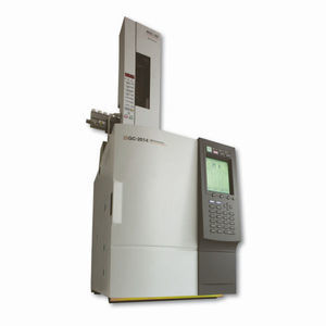
Gas Chromatograph
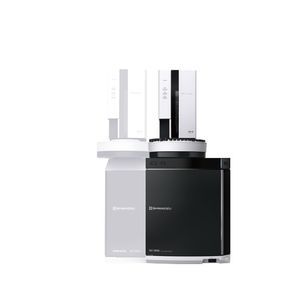
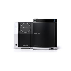
Gas Chromatograph-Mass Spectrometry
Columns, Reagents and Consumables

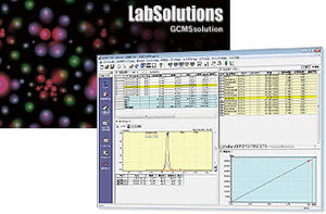
Software & Informatics


MALDI-Based Instruments and Solutions

Molecular Spectroscopy


Elemental Analysis

Life Science Lab Instruments

Material Testing

Total Organic Carbon Analysis

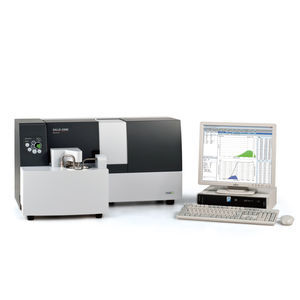
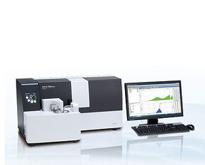
Particle Size Analysis

Remove all
Compare up to 10 products