
{{product.productLabel}} {{product.model}}
{{#if product.featureValues}}{{product.productPrice.formattedPrice}} {{#if product.productPrice.priceType === "PRICE_RANGE" }} - {{product.productPrice.formattedPriceMax}} {{/if}}
{{#each product.specData:i}}
{{name}}: {{value}}
{{#i!=(product.specData.length-1)}}
{{/end}}
{{/each}}
{{{product.idpText}}}

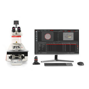
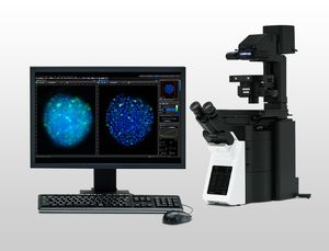
... & 3D spatial relational analysis for objects of any type, morphological complexity, and object hierarchy. Advanced data analysis accessible for all Quickly train laboratory users on the platform, ...
Leica Microsystems

... standards to customize the analysis to organizational norms and processes, and quickly update any official standard changes Optimize your workflow Achieve a rapid workflow for inclusion analysis. ...
Leica Microsystems
... analyzing particle quantity, size, and composition. Cleanliness analysis systems from Leica Microsystems offer optimized software and a unique 2-in-1 solution for visual and chemical analysis. Your ...
Leica Microsystems
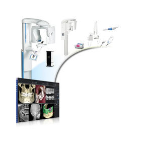
... standard formats, such as JPEG, DICOM and STL, or to launch data directly into 3rd party software. Practice management integration is easy to establish with over 100 software options. Superior usability Romexis ...
Planmeca
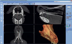
... intuitive imaging software with features and functionalities designed especially for veterinary needs. It lets veterinarians enjoy all the benefits of digital dentistry – with just one software. All-in-one ...
Planmeca

The Planmeca Romexis® Cephalometric Analysis software module is the ultimate tool for performing cephalometric analyses, surgical planning and treatment follow-ups in 2D. The software ...
Planmeca
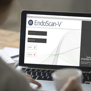
... quality control manager’s top priority. Endosafe® EndoScan-V™ is our foundational and secure endotoxin measuring and analysis software for generating and reporting quantitative LAL and rCR test data. Establish ...

... first time. Charles River Cortex is an all-encompassing endotoxin software platform for data and instrument management, investigation analysis, and process monitoring. This software ...

... comparison against our best-in-industry Accugenix® sequence libraries. Our expert phylogenetic data analysis team will review the data and provide their analysis and interpretation in a comprehensive ...
Control software - eControl eControl for your NEXOPART Air Jet Sieving Machine e200 LSThe eControl software is already integrated into each Air Jet Sieving Machine e200 LS. You can choose from a selection ...
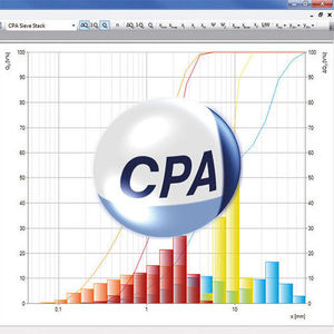
CpaServ. Modern, convenient, accessible.The NEXOPART CpaServ software is backed by the artificial intelligence of the CPA 2-1 analyzer. CpaServ offers you maximum ease of use, a modern user interface and provides automated ...
... Cardiovascular Surgery). Suitestensa consolidates all clinical and administrative information into a single user interface, serving as the central hub for collecting and distributing data, whether it be clinical ...
Esaote
... radiologist. CAAS MRV (Magnetic Resonance Ventricular analysis software) and CAAS MR Flow (Magnetic Resonance Flow analysis software) help you by providing the relevant ...
Esaote

... Quantitative X-Ray Angiography Software The gold standard in Quantitative Analysis Software The gold standard CAAS platform represents the widest range of post-processing images solutions ...
Esaote
... image acquisition, powerful analysis tools, and so much more. Dimension MultipositionCount & MeasureCI DeconvolutionWell Plate NavigatorRatio / FRETPhoto ManipulationDatabase CoreDatabase ClientNetCamLife Science ...
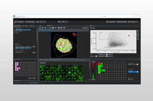
Insightful Analysis, Intelligent Answers NoviSight 3D cell analysis software advances your discovery by providing statistical data for spheroids and other 3D objects in microplate-based ...

Neowise is an innovative software designed to optimise daily radiological practice, integrating advanced features that simplify both clinical activities and communication. Discover how Neowise can transform ...

... heart of the iQue® platform, iQue Forecyt® Software, is integrated, interactive assay development, implementation, and analysis visual environment. Designed for speed to actionable answers, iQue Forecyt® ...
Sartorius Group

Leverage Workcloud Sync for synchronous communication, allowing real-time exchanges and different forms of communication to empower frontline workers. Our communication tools help create a more engaged, collaborative, and productive workforce.
... efficiency, functionality, and the flow of clinical information with Intesys® Clinical Suite. Our clinical system carries vital information to clinicians across services and sites. ICS ...
Spacelabs Healthcare
... Holter analysis while working from home using a dedicated installation of Pathfinder SL supplied on a laptop PC. Continue to diagnose your patients quickly and efficiently while removing the need to be on site. Developed ...
Spacelabs Healthcare
The Spacelabs Lifescreen Pro Event Screening System is key to a high diagnostic yield for patients with suspected or intermittent arrhythmias, providing fast, assisted analysis when and where it’s needed ...
Spacelabs Healthcare

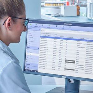
INSTINCT S is Hamilton's powerful, easy-to-use lab sample management software that allows users to run daily operations with minimal training. The sample management software is available across all Hamilton ...

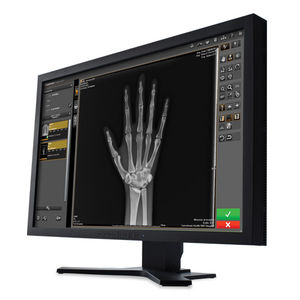
... every image and that the software can be used intuitively and logically. We attach special importance to perfectly supporting the efficiency of DÜRR MEDICAL systems, be it CR or DR Why DÜRR MEDICAL ...

W20s is a software that records the signals obtained through the SIBELMED spirometer, so that they can later be analyzed and saved in a simple and intuitive way. It is compatible with the DATOSPIR range, and can operate ...
SIBELMED

SIBELMED W50 is a software for viewing, storing, transferring, analyzing and introducing audiometric tests, compatible with the range of SIBELMED audiometers. Audiometric charts With the test results Different ...
SIBELMED

... Windows operating system VISTA, 7, 8.1 and 10 Manual or automatic events analysis The technician can manually mark the events or use the automatic analysis function of the software, ...
SIBELMED
LabX™ Balance software integrates fully with laboratory information systems, such as LIMS and chromatography software, and enables a seamless flow of data throughout the entire analysis ...

... Connectivity Connect SevenCompactTM, SevenCompactTM Duo, Seven2GoTM Pro, FiveEasyPlus or SevenExcellenceTM to EasyDirectTM pH software either by USB or RS232 connection. Up to three instruments can be connected simultaneously ...
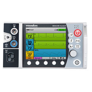
... debriefing gives everyone the opportunity to talk about a stressful situation and to improve patient care processes. DEFIview software helps you to display session data recorded by MEDUCORE Standard². If you analyze reactions ...

... downloadable ImageScope viewing software—experience rapid access to crisp, true-color digital slide images to which you can adjust magnification, pan and zoom, compare different stains, annotate areas of interest, perform ...
... data storage Product Details CLC Genomics Server offers a unique and stable software architecture core that makes it possible to apply a range of bioinformatics analysis services on high-throughput ...
QIAGEN

... years of computerised CTG analysis, Sonicaid FetalCare 3 software is an essential tool for modern obstetric management. Running on a PC, it receives data from the fetal monitor via a cable connection. Its ...

... developed) and therefore will not perform in the same way. WHAT IS DAWES-REDMAN CTG ANALYSIS? It is a unique software tool which provides a numeric analysis of the CTG trace and ...

... offer a range of software packages for Life and Physical science research. Solis – Camera and Spectroscopy Control Software SDK Library – Data Acquisition Configuration Software GPU ...

... the stress. MassLynx Quantitation Applications Perform quantitative mass spectrometry analysis with MassLynx Quantitation Applications, mass spec software offerings include TargetLynx XS, Quanpedia, ...
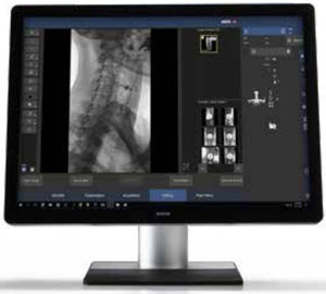
... Learning algorithm automatically adjusts images as they should be presented to the radiologist Dose Management through Diagnostic Reference Levels (DRL) and Extended Dose Reporting Cyber-Security & Encryption: Transport ...
... acquisition and management for CR and DR Systems, Image Suite V4 Software has set a new benchmark for workflow efficiency in smaller facilities. Powered by our Eclipse Engine, this robust software offers: An ...
Carestream

The SUREengine is a real-time, high-speed image processing engine that increases the sharpness, contrast, and graininess of fluoroscopy and radiography images and simultaneously increases operation speed to support quicker treatments.
Shimadzu Europe Medical Systems
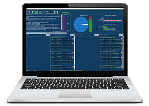
... Gain peace of mind in knowing that your analyzers are performing to the right standards. Peer comparison makes a statistical analysis of mean bias value – an analyzer’s bias from a target value – compared to analyzers ...
... in your daily activities Get the most from your clinical cases Whatever your specialty, AIS is the ideal companion for your diagnostics, providing all you need to get a reliable patient clinical ...
Please specify:
Help us improve:
remaining