- Primary care
- General practice
- Body anatomical model
- Shandong Digihuman Technology CO., Inc.
- Products
- Catalogs
- News & Trends
- Exhibitions
Body anatomical model for teachingembryoresin

Add to favorites
Compare this product
fo_shop_gate_exact_title
Characteristics
- Area of the body
- body
- Procedure
- for teaching
- Type
- embryo
- Material
- resin
- Color
- pink
- Length
34 cm
(13.4 in)- Weight
750 g
(26.46 oz)
Description
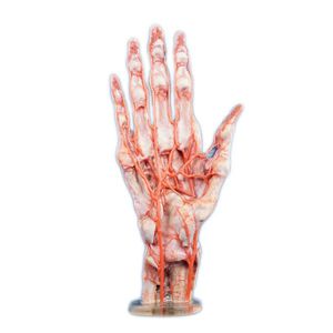
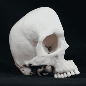
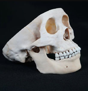
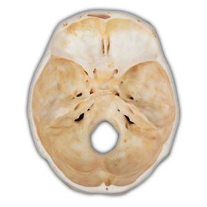
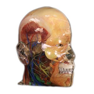
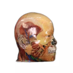
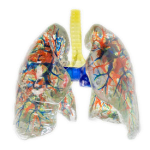
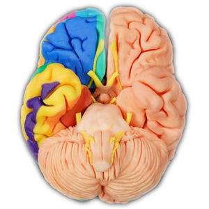
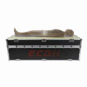
Constructing a multi-structured 3D digital anatomical model from high-precision digitized human body data. Full-color, multi-material 3D printer and environmentally friendly resin material printing, providing 1:1 high simulation of physical anatomical models.
Cloacal Malformation Combined with Polycystic Kidney
Week:13 weeks
no anus, 0.7*0.2 cm protrusion visible at the external genitalia, segmental thickened intestinal canal below the appendix, about 1.0 cm long.
The left kidney showed polycystic-like changes, with slender ureter (atresia) and dilated ureter externally.
The left extra-renal pelvis was dilated, the ureter was not accessible, and there was no appendix. There was no response at the external genitalia by pumping water into the bladder; the left kidney was in a low position and the intestinal canal was about 40 cm long. (iv) Cloacal malformation with complete urorectal diaphragm sequence sign.
Dewlap with Cleft Lip and Palate
Week: 12.4 weeks
Dewlap malformation, single umbilical artery, right cleft lip III°, size 0.3*0.2 cm.
Cleft alveolar arch, complete cleft palate.
amniotic band adhesions were seen in the 3rd and 4th fingers of the left hand (fingers separated), in the 2nd, 3rd and 4th fingers of the right hand (parallel fingers), in the 1st, 2nd and 3rd toes of the left foot (parallel fingers and short toes), and in the 1st toe of the right foot.
Umbilical Bulge
Week:23.2 weeks
Fetal umbilical pang out (the bulge is intestinal tube, liver and appendix), size 5*4.5*2.5cm.
X-ray showed that the 6th, 7th and 10th vertebrae were dysplastic.
VIDEO
Catalogs
No catalogs are available for this product.
See all of Shandong Digihuman Technology CO., Inc.‘s catalogsExhibitions
Meet this supplier at the following exhibition(s):


Other Shandong Digihuman Technology CO., Inc. products
3D Printing Model
Related Searches
- Anatomy model
- Demonstration anatomical model
- Teaching anatomy model
- Intracranial anatomical model
- Transparent anatomical model
- Whole body anatomical model
- White anatomical model
- Nervous system model
- Facial model
- Artery model
- Anatomical model with nerves
- Respiratory tract anatomical model
- Anatomical model with musculature
- Cerebral anatomical model
- Anatomical model with blood vessels
- Pink anatomical model
- Lung model
- Resin anatomical model
- Vein model
- Hand anatomical model
*Prices are pre-tax. They exclude delivery charges and customs duties and do not include additional charges for installation or activation options. Prices are indicative only and may vary by country, with changes to the cost of raw materials and exchange rates.


