- Primary care
- General practice
- Hand anatomical model
- Shandong Digihuman Technology CO., Inc.
- Products
- Catalogs
- News & Trends
- Exhibitions
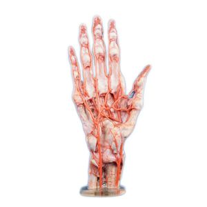
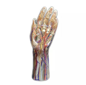
Hand anatomical model for teachingresinwith musculature
Add to favorites
Compare this product
fo_shop_gate_exact_title
Characteristics
- Area of the body
- hand
- Procedure
- for teaching
- Material
- resin
- Accessories
- with nerves, with musculature, with blood vessels
- Other characteristics
- transparent
Description
Constructing a multi-structured 3D digital anatomical model from high-precision digitized human body data. Full-color, multi-material 3D printer and environmentally friendly resin material printing, providing 1:1 high simulation of physical anatomical models.
Data Source: Digital Human Model
The 3D printed model of hand blood vessels and nerves is a transparent package model. The skin, superficial fascia, deep fascia, and other structures are all transparent materials, and the bones, ligaments, blood vessels, nerves, and muscles are full-color materials. Observe the internal anatomy through the transparent materials. The internal anatomical structure is consistent with the real specimen, and the transparent wrapping material maintains the spatial positional relationship of the anatomical structure, and can truly express the morphological characteristics of normal hand blood vessels, nerves, and muscles
Data Source
Data were selected from a high-precision digital human dataset, including raw tomographic data, refined segmentation data and 3D geometric models of organ structures, including bone, muscle, blood vessels, nerves, ligaments, and other anatomical structures, and the voxel size of this example dataset was 0.0384mm*0.0384mm*0.1mm. Also, cadaveric specimens fixed by formalin were selected as a 3D printing the basis for reference and comparison of the model.
Modeling: The voxels of each anatomical structure surface were extracted from the original tomographic dataset to generate texture maps of the geometric model to ensure that the appearance of the geometric model of each anatomical structure has the same visual perception as that of the real anatomical specimen
Catalogs
No catalogs are available for this product.
See all of Shandong Digihuman Technology CO., Inc.‘s catalogsExhibitions
Meet this supplier at the following exhibition(s):


Other Shandong Digihuman Technology CO., Inc. products
3D Printing Model
Related Searches
- Anatomy model
- Demonstration anatomical model
- Teaching anatomy model
- Intracranial anatomical model
- Transparent anatomical model
- Whole body anatomical model
- White anatomical model
- Nervous system model
- Facial model
- Artery model
- Anatomical model with nerves
- Respiratory tract anatomical model
- Anatomical model with musculature
- Cerebral anatomical model
- Anatomical model with blood vessels
- Pink anatomical model
- Lung model
- Resin anatomical model
- Vein model
- Hand anatomical model
*Prices are pre-tax. They exclude delivery charges and customs duties and do not include additional charges for installation or activation options. Prices are indicative only and may vary by country, with changes to the cost of raw materials and exchange rates.


