- Primary care
- General practice
- Skull model
- Shandong Digihuman Technology CO., Inc.
- Products
- Catalogs
- News & Trends
- Exhibitions
Skull model for teachingwhite
Add to favorites
Compare this product
fo_shop_gate_exact_title
Characteristics
- Area of the body
- skull
- Procedure
- for teaching
- Color
- white
Description
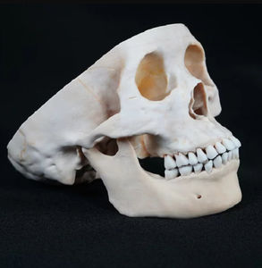
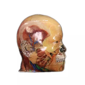
A printed model of the mid-sagittal section of the skull, accurately replicating its internal and external anatomical features using high-precision digital human data, serves as a realistic substitute for studying skull anatomy.
Data Source: Digital Human Model
The mid-sagittal section of the skull is a printed model of the mid-sagittal section of the skull, showing the internal and external views of the skull. This model has the same visual perception and touch as the real mid-sagittal section of the skull. It can truly display the internal and external anatomical features of the skull, such as the bony protrusions on the outer wall of the bony nasal cavity, the opening of the sinuses and other complex anatomy The structure can be used as a substitute for the sagittal section of the skull.
Data Resources
Data Source: select data from the high-precision digital human data set, including original sectional data, refined segmentation data, and three-dimensional geometric model of organ structure, including anatomical structures such as bone, muscle, blood vessel, nerve, and ligament. The voxel size of this data set is 0.0384mm * 0.0384mm * 0.1mm. At the same time, the cadaveric specimens fixed with formalin are selected as the reference and comparison basis of the 3D printing model.
Model Making: the voxels in the volume data on the surface of each anatomical structure are extracted from the original section data set, and the texture map of the geometric model is generated to ensure that the appearance of the geometric model of each anatomical structure has a visual perception consistent with the real anatomical specimen.
Catalogs
No catalogs are available for this product.
See all of Shandong Digihuman Technology CO., Inc.‘s catalogsExhibitions
Meet this supplier at the following exhibition(s):


Other Shandong Digihuman Technology CO., Inc. products
3D Printing Model
Related Searches
- Anatomy model
- Demonstration anatomical model
- Teaching anatomy model
- Intracranial anatomical model
- Transparent anatomical model
- Whole body anatomical model
- White anatomical model
- Nervous system model
- Facial model
- Artery model
- Anatomical model with nerves
- Respiratory tract anatomical model
- Anatomical model with musculature
- Cerebral anatomical model
- Anatomical model with blood vessels
- Pink anatomical model
- Lung model
- Resin anatomical model
- Vein model
- Hand anatomical model
*Prices are pre-tax. They exclude delivery charges and customs duties and do not include additional charges for installation or activation options. Prices are indicative only and may vary by country, with changes to the cost of raw materials and exchange rates.


