- Primary care
- General practice
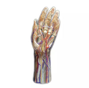
- Body anatomical model
- Shandong Digihuman Technology CO., Inc.
- Products
- Catalogs
- News & Trends
- Exhibitions
Body anatomical model for teachingembryoresin
Add to favorites
Compare this product
fo_shop_gate_exact_title
Characteristics
- Area of the body
- body
- Procedure
- for teaching
- Type
- embryo
- Material
- resin
- Color
- pink
- Length
Max.: 270 mm
(10.6 in)Min.: 2 mm
(0.1 in)
Description
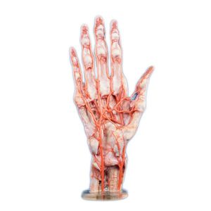
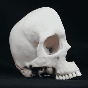
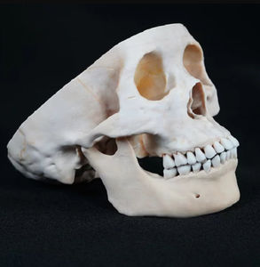
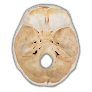
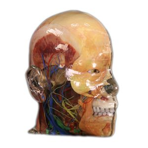
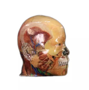
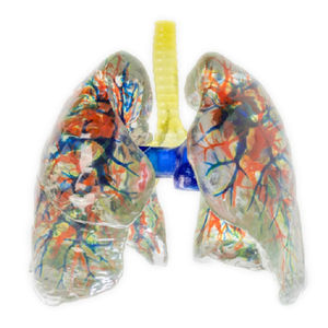
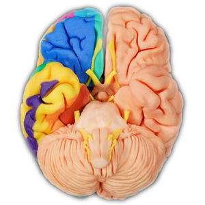
Constructing a multi-structured 3D digital anatomical model from high-precision digitized human body data. Full-color, multi-material 3D printer and environmentally friendly resin material printing, providing 1:1 high simulation of physical anatomical models.
Early embryonic stage (1-5 days) Ratio:200:1
1 day old embryo: fertilized egg with outer hyaline band. Blastocyst size: 0.2 mm 2-day-old embryo: 2-cell stage, consisting of two ovoid spheres with outer zona pellucida and 2 to 3 polar bodies visible. Blastocyst size: 0.2mm 3-day-old embryo: mulberry embryo, composed of about 16 ovoid spheres, resembling a mulberry, and covered with a zona pellucida. Blastocyst size: 0.2mm 4-day-old embryo: blastocyst, also known as blastocyst, with the trophectoderm surrounding the blastocyst cavity, with a cluster of inner cells at one end of the cavity and a zona pellucida around the blastocyst.
Early embryonic stage (6-14 days) Ratio:50:1
6 days old embryo: onset of implantation: adhesion of the trophoblast end of the blastocyst to the endometrium. Embryo blastocyst size: 0.25mm 7-day-old embryo: embryo in implantation, blastocyst partially enters the endometrium and the trophectoderm differentiates into the cytotrophectoderm and the syncytial trophectoderm. Blastocyst size: 0.25mm 9-day-old embryo: The blastocyst penetrates deeply into the endometrium, the implantation opening is sealed by the coagulation plug, the dictyostelium is formed, the amniotic sac and primary yolk sac appear one after another, and the trophectoderm traps appear in the syncytial trophectoderm.
Catalogs
No catalogs are available for this product.
See all of Shandong Digihuman Technology CO., Inc.‘s catalogsExhibitions
Meet this supplier at the following exhibition(s):


Other Shandong Digihuman Technology CO., Inc. products
3D Printing Model
Related Searches
- Anatomy model
- Demonstration anatomical model
- Teaching anatomy model
- Intracranial anatomical model
- Transparent anatomical model
- Whole body anatomical model
- White anatomical model
- Nervous system model
- Facial model
- Artery model
- Anatomical model with nerves
- Respiratory tract anatomical model
- Anatomical model with musculature
- Cerebral anatomical model
- Anatomical model with blood vessels
- Pink anatomical model
- Lung model
- Resin anatomical model
- Vein model
- Hand anatomical model
*Prices are pre-tax. They exclude delivery charges and customs duties and do not include additional charges for installation or activation options. Prices are indicative only and may vary by country, with changes to the cost of raw materials and exchange rates.


