
{{product.productLabel}} {{product.model}}
{{#if product.featureValues}}{{product.productPrice.formattedPrice}} {{#if product.productPrice.priceType === "PRICE_RANGE" }} - {{product.productPrice.formattedPriceMax}} {{/if}}
{{#each product.specData:i}}
{{name}}: {{value}}
{{#i!=(product.specData.length-1)}}
{{/end}}
{{/each}}
{{{product.idpText}}}
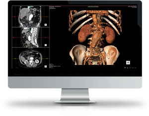
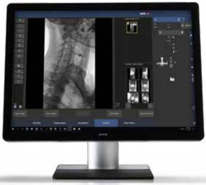
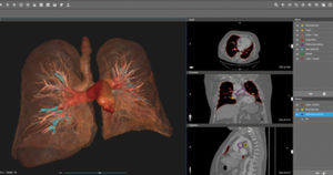
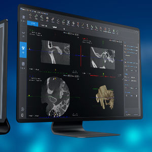
... including radiographic imaging and computed tomography including cone-beam CT reconstruction tools Enhance your workflows with the best computer-aided software analysis tools Multi-disciplinary collaboration ...
VAREX Imaging

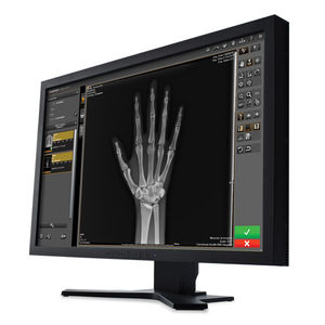
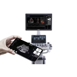
... to install any software. Whether you work for a small diagnostic center or for a complex diagnostic imaging network, ZEfiRO adapts seamlessly to meet your needs, leveraging ...
Esaote
... Radiology, transforming the diagnostic process from patient admission to the distribution of clinical data outside the Department and Hospital. • Pure Web Technology. • Patient file and complete ...
Esaote
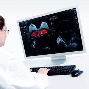
SUITESTENSA MG - Mammography Information System and PACS • Breast Cancer Screening Programs in integrated HIS, RIS PACS environment. • Double-blind reading protocols, review process with automatic arbitration and reassignment ...
Esaote
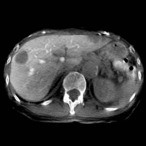
This function is for observing cross-sectional images of low-contrast regions, primarily tumor stains during procedures.
Shimadzu Europe Medical Systems
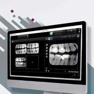
... reliable patient clinical landscape, to view, process and merge dental imaging data and boost treatment acceptance with diagnostic reporting tools. A perfect companion for your practice AIS ...

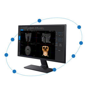
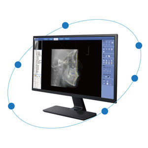
Neowise is an innovative software designed to optimise daily radiological practice, integrating advanced features that simplify both clinical activities and communication. Discover how Neowise can transform ...
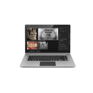
... STL, or to launch data directly into 3rd party software. Practice management integration is easy to establish with over 100 software options. Superior usability Romexis has been designed for the ...
... Romexis® software platform offers versatile and intuitive tools for varying clinicians and specialities – helping dental clinics of all types reach their full potential and provide the best level of care possible. The ...

... for viewing 2D and 3D images captured with Planmeca Group’s imaging devices. The application can be exported and sent together with the veterinary dental X-rays from the Planmeca Romexis® software to ...
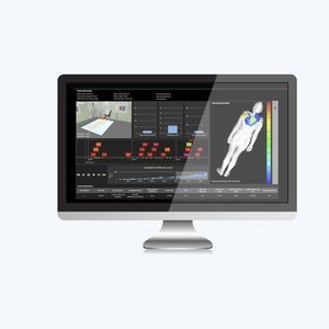
... Map DoseWatch provides an interactive tool to support the post-procedure review conducted by the interventional imaging physician or medical physicist as they perform their comprehensive assessment of ...
GE Healthcare
... Learning algorithm automatically adjusts images as they should be presented to the radiologist Dose Management through Diagnostic Reference Levels (DRL) and Extended Dose Reporting Cyber-Security & Encryption: Transport ...
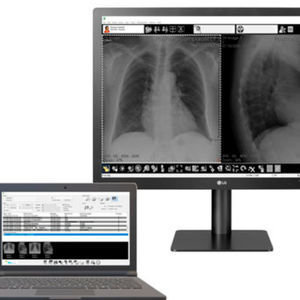
... acquisition and management for CR and DR Systems, Image Suite V4 Software has set a new benchmark for workflow efficiency in smaller facilities. Powered by our Eclipse Engine, this robust software offers: An ...
Carestream
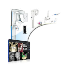
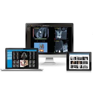
Discover a new generation of dental imaging software with CS Imaging version 8—the platform that provides one-stop access to all your 2D images, 3D images and CAD/CAM data—to manage your ...
Carestream Dental
... produce an X-ray software that promotes clinical efficiency and usability so your patients leave your practice with a healthy and happy smile. Sidexis 4 provides you with the tools you need to help you ...
... exported from the D2P software can be used in a wide variety of applications including 3D printers, VR devices, surgical planning software, and CAD software. D Systems is the only manufacturer ...
3D Systems
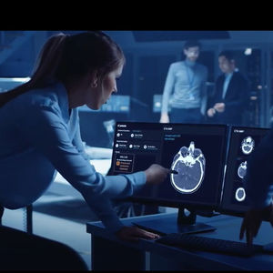
... intelligence to our Collaborative imaging offering, Automation Platform is an AI-based, zero-click solution that uses deep learning technology to streamline your workflow for fast, actionable results every time. From ...
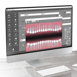
... practice computers or via the iRYS Viewer application available for iPAD. Dose Book Tool A feature designed to make iRYS software more efficient at gathering and managing information on acquired scans: the system ...
... uninterrupted workflows. It supports standard protocols such as DICOM for medical imaging and also provides several tools that allow advanced customisation. Customised user profiles The software ...
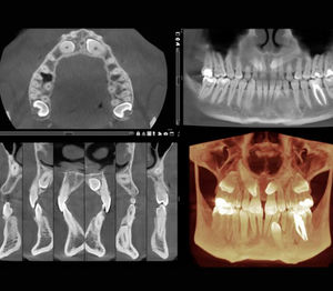
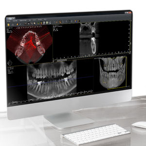
Taking a 3D cone beam image with I-Max 3D or I-Max Ceph 3D. The 12x10mm FOV provides a complete clinical view of the patient, ideal for implant surgery treatments. Digital Impressions with Owandy IOS. The integration ...
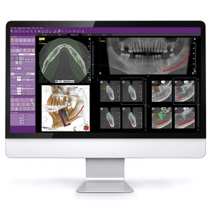
QuickVision imaging software is especially designed for dental surgeries. It includes a patient database, an imaging module and a dental diagram. Key features 1 - Patient database Simple ...
... This includes the precise location of the implant, potential collisions, and various other clinical aspects. The QuickVision 3D implant planning software will be your greatest ally in achieving a ...
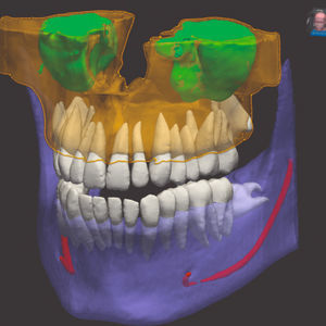
... metal artifact removal, the system corrects metal artifacts intelligently. It avoids overmodification and saves the original clinical data. Implant Simulation The bone and bone mass in the implant area will be evaluated ...
... Measurement Analysis Methods There are 19 measurement methods built into the software, which can be selected by doctors according to the actual clinical situation. Meanwhile, the software ...
... area of the airway in the form of chromatographic visualization. Medical Images Comparison By loading multiple images, it is possible to perform single-slice comparison medical imaging. ...
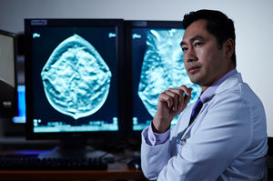
Create comprehensive diagnostic studies that reveal exceptional clinical detail and sensitivity with Contrast Enhanced Mammography 2D imaging (CEM). Contrast Enhanced 2D imaging ...
Hologic

... HIPAA-compliant to provide a secure and trusted platform for medical professionals. Anatomage brings it's award-winning Invivo 3D imaging technology to the cloud in the form of a zero-footprint 3D ...
Anatomage
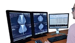
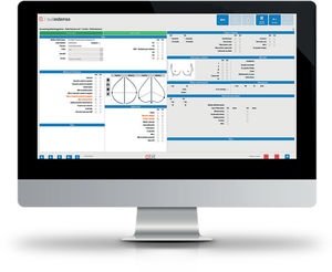
Med Mammo is a mammography diagnostic workstation which combines ease of use and high performance features. Med Mammo allows multimodality image reading including tomosynthesis Interoperability and constructors' ...
Medecom
... Does not need extra hardware The stitching module is compatible to: -> Our acquisition workstation : Med-XRay -> Our diagnostic workstation : Med-Diag
Medecom
Med Diag is a multimodality workstation which main purpose is to print in a very efficient way. Med Diag We give the user all the tools to combine speed and high performance features. CR and DR workstation used by some of the leaders. Simple ...
Medecom

Scatter correction software increases the contrast of images taken without a grid up to a level similar to images taken with a grid. Applicable to: Arcoma Intuition and Precision.

... tools and sophisticated archiving functionality. BRAIN QUICK EEG software is adaptable to any size facility, from large hospitals to private practice and research centers. Software Key Features: SQL ...

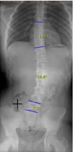
... navigate menus and tools. IMAGING AND ANNOTATION TOOLS Advanced tools include: Smart Window Level, Crop, Mask, Heart-to-Lung Ratio, Center Line, Cobb Angle, Image Tile-View, and more. DICOM & HL7 COMPLIANT XC's ...
... in and click "Product Enquiry" below, leave your message and we will contact you in time. Online Solution Kit for Medical Imaging In the days of COVID-19, webinars have given us a safe way to keep ...

NEOWISE is an innovative software designed to assist doctors in their daily practice, integrating advanced features to streamline clinical and communication activities. Single interface Centralised ...
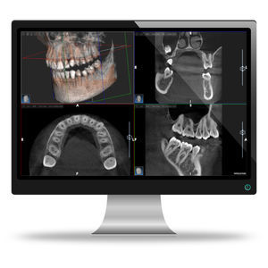
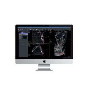
... thanks to the advanced systems adopted by NewTom. Increasingly straightforward and efficient operational flow and clinical and diagnostic activities. Complete view of the dental arches in cross sections ...
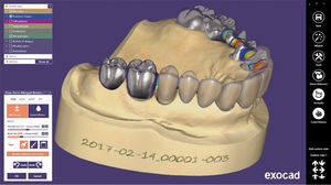
exocad Your future in digital dentistry exocad offers complete software solution for digital dentistry DENTALCAD Powerful dental CAD software exocad CAD software is known for its ...
Please specify:
Help us improve:
remaining