
- Laboratory
- Laboratory medicine
- Harris hematoxylin reagent
- Wuhan Jinhong Biotech Development Co., Ltd.
- Company
- Products
- Catalogs
- News & Trends
- Exhibitions
Harris hematoxylin reagent kit H-E stainfor histopathology

Add to favorites
Compare this product
Characteristics
- Type
- Harris hematoxylin
- Applications
- for histopathology
Description
It is mainly intended to display the common morphological structure for normal components of various tissues and pathological changes. H-E stain is the basic and necessary method in biology, histology, pathology and cytology. It is widely used in diagnostics, teaching and research, with important values.
Principle:
The nucleolus, which is made up of acid materials, has a strong affinity for alkaline stain (Hematoxylin) while the cytoplasm, made up of alkaline materials, binds favorably to acid stain (Eosin). Therefore, in the presence of the H-E stain, nucleolus will be stained bright indigo blue by hematoxylin; cytoplasm, stroma, muscle fiber and collagen fiber will be varying shades of pink; and erythrocyte will be salmon pink.
Methods:
1. Deparaffinize tissue in two washes of xylene for 5 minutes each. Wash tissue twice in 95% ethanol for 1 minute each.
2. Place tissue in 80% ethanol for 1 minute, then rinse with tap water for 1 minute.
3. Stain for 3~5 minutes with Harris. Rinse with tap water for 1~2 minutes.
4. Place tissue in 0.5%~1% hydrochloric acid ethanol for a few seconds.
5. Wash the 'Blue' tissue in tap water, or with lithium carbonate solution, for 5~10 minutes, followed by another water rinse for 1~2 minutes.
6. Place tissue in Eosin for 30~60 seconds. Rinse with water.
7. Dehydrate tissue in 80% and 95% ethanol, respectively.
8. Dehydrate in 100% ethanol for 1~2 minutes, then clear in phenol xylene and xylene for 1~2 minutes each.
9. Mount with mounting media and examine microscopically.
Catalogs
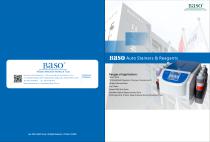
Catalogue of Baso
16 Pages
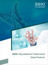
Mycobaterium Tuberculosis Series
20 Pages
Other Wuhan Jinhong Biotech Development Co., Ltd. products
Pathohistology Stains


Related Searches
- Baso test kit
- Baso solution reagent
- Blood assay kit
- Molecular biology reagent kit
- Serum assay kit
- Plasma assay kit
- Blood rapid diagnostic test
- Baso research reagent
- Baso laboratory reagent
- Baso diagnostic reagent
- Baso histology reagent
- Whole blood rapid diagnostic test
- Clinical assay kit
- Baso cytology reagent
- Baso stain reagent
- Clinical chemistry reagent
- Reagent medium reagent kit
- Baso bacteria reagent
- Blood sample reagent kit
- Baso clinical reagent
*Prices are pre-tax. They exclude delivery charges and customs duties and do not include additional charges for installation or activation options. Prices are indicative only and may vary by country, with changes to the cost of raw materials and exchange rates.