- Laboratory
- Laboratory medicine
- Staining solution reagent
- BIO-OPTICA Milano
- Company
- Products
- Catalogs
- News & Trends
- Exhibitions
Staining solution reagent May Grünwald Giemsafor histologyfor cytology
Add to favorites
Compare this product
fo_shop_gate_exact_title
Characteristics
- Type
- staining solution
- Applications
- for histology, for cytology
Description
Minimum number of tests that can be performed 100
Completion time 35 minutes
Shelf life 2 years
Storage conditions 15-25°C
Additional equipment Graduated cylinder
Application
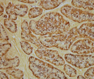
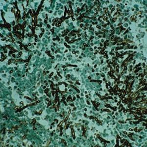
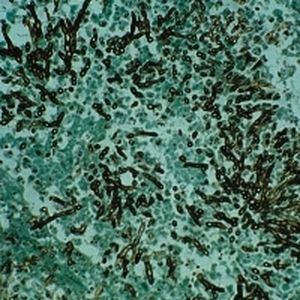
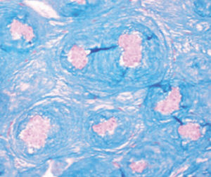
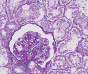
The method of choice for differentiating cell types and highlighting parasites on tissue
sections; particularly indicated for lymphohematopoietic tissue. This stain is often used
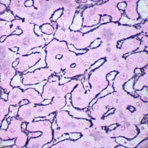
for identifying endothelial reticulum.
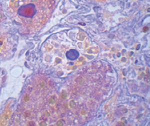
Result
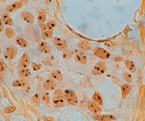
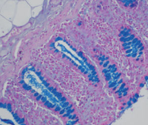
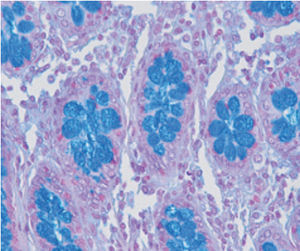
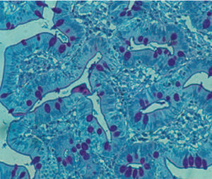
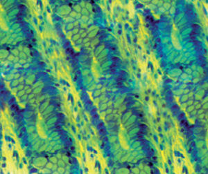
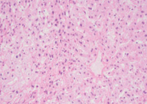
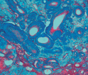
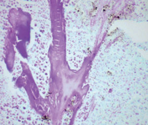
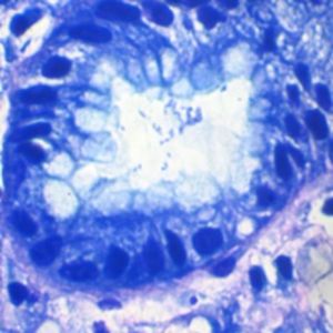
Nuclei blue
Basophilic cytoplasm from sky blue to dark blue
Acidophilic cytoplast pink
Bacteria blue
Product for the préparation of cyto-histological samples for optical microscopy.
Recommended method to differentiate cell types and to reveal parasites in tissue sections. Especially useful for lymphopoietic tissue. This stain is often used to demonstrate endothélial réticulum.
PRINCIPLE
- - May Grunwald solution, consisting of eosin-methylene blue, stains nudei blue and basophil cytoplasm pinkish red.
- - Giemsa solution, a complex consisting of methylene blue chloride, eosin-methylene blue and azuré II eosinate, improves the intensity of nuclear staining and the capacity to show selectively cellular structures.
To appreciate results always remember two factors: pH of washing water and dilution buffer hâve a strong influence on final colour chart; intensity of stain may vary according to différentiation time.
Catalogs
General Catalogue
139 Pages
Related Searches
- Bio-Optica solution reagent
- Laboratory reagent kit
- Bio-Optica histology reagent
- Reagent medium reagent kit
- Bio-Optica stain reagent
- Bio-Optica cytology reagent
- Buffer solution reagent kit
- Bacteria reagent kit
- Bio-Optica staining solution reagent
- Microscope slide
- Sample preparation reagent kit
- Pathology reagent
- Bilirubin reagent kit
- Bio-Optica fixative solution reagent
- Paraffin wax reagent
- Phosphate buffer reagent kit
- Collagen reagent kit
- Helicobacter pylori reagent kit
- Microscopy reagent
- Decalcifying solution reagent
*Prices are pre-tax. They exclude delivery charges and customs duties and do not include additional charges for installation or activation options. Prices are indicative only and may vary by country, with changes to the cost of raw materials and exchange rates.


